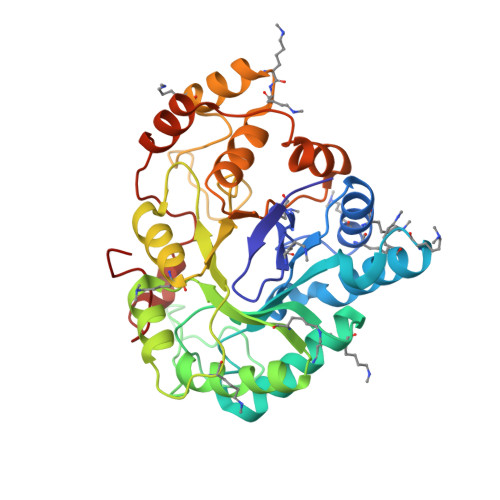
4ICC

Crystal structure of human AKR1B10 complexed with NADP+ and JF0064


Experimental Data Snapshot

  • Method: X-RAY DIFFRACTION
  • Resolution: 1.75 Å
  • R-Value Free: 0.186 
  • R-Value Work: 0.162 
  • R-Value Observed: 0.163 

Starting Model: experimental
View more details

wwPDB Validation   3D Report Full Report


Ligand Structure Quality Assessment 


This is version 1.3 of the entry. See complete history


Literature

Identification of a novel polyfluorinated compound as a lead to inhibit the human enzymes aldose reductase and AKR1B10: structure determination of both ternary complexes and implications for drug design.

Cousido-Siah, A.Ruiz, F.X.Mitschler, A.Porte, S.de Lera, A.R.Martin, M.J.Manzanaro, S.de la Fuente, J.A.Terwesten, F.Betz, M.Klebe, G.Farres, J.Pares, X.Podjarny, A.

(2014) Acta Crystallogr D Biol Crystallogr 70: 889-903

  • DOI: https://doi.org/10.1107/S1399004713033452
  • Primary Citation of Related Structures:  
    4ICC, 4IGS

  • PubMed Abstract: 

    Aldo-keto reductases (AKRs) are mostly monomeric enzymes which fold into a highly conserved (α/β)8 barrel, while their substrate specificity and inhibitor selectivity are determined by interaction with residues located in three highly variable external loops. The closely related human enzymes aldose reductase (AR or AKR1B1) and AKR1B10 are of biomedical interest because of their involvement in secondary diabetic complications (AR) and in cancer, e.g. hepatocellular carcinoma and smoking-related lung cancer (AKR1B10). After characterization of the IC50 values of both AKRs with a series of polyhalogenated compounds, 2,2',3,3',5,5',6,6'-octafluoro-4,4'-biphenyldiol (JF0064) was identified as a lead inhibitor of both enzymes with a new scaffold (a 1,1'-biphenyl-4,4'-diol). An ultrahigh-resolution X-ray structure of the AR-NADP(+)-JF0064 complex has been determined at 0.85 Å resolution, allowing it to be observed that JF0064 interacts with the catalytic residue Tyr48 through a negatively charged hydroxyl group (i.e. the acidic phenol). The non-competitive inhibition pattern observed for JF0064 with both enzymes suggests that this acidic hydroxyl group is also present in the case of AKR1B10. Moreover, the combination of surface lysine methylation and the introduction of K125R and V301L mutations enabled the determination of the X-ray crystallographic structure of the corresponding AKR1B10-NADP(+)-JF0064 complex. Comparison of the two structures has unveiled some important hints for subsequent structure-based drug-design efforts.


  • Organizational Affiliation

    Department of Integrative Biology, Institut de Génétique et de Biologie Moléculaire et Cellulaire, CNRS/INSER/UdS, 1 Rue Laurent Fries, 67404 Illkirch CEDEX, France.


Macromolecules
Find similar proteins by:  (by identity cutoff)  |  3D Structure
Entity ID: 1
MoleculeChains Sequence LengthOrganismDetailsImage
Aldo-keto reductase family 1 member B10A [auth X]316Homo sapiensMutation(s): 2 
Gene Names: AKR1B10AKR1B11
EC: 1.1.1 (PDB Primary Data), 1.1.1.300 (UniProt), 1.1.1.54 (UniProt)
UniProt & NIH Common Fund Data Resources
Find proteins for O60218 (Homo sapiens)
Explore O60218 
Go to UniProtKB:  O60218
PHAROS:  O60218
GTEx:  ENSG00000198074 
Entity Groups  
Sequence Clusters30% Identity50% Identity70% Identity90% Identity95% Identity100% Identity
UniProt GroupO60218
Sequence Annotations
Expand
  • Reference Sequence
Small Molecules
Ligands 2 Unique
IDChains Name / Formula / InChI Key2D Diagram3D Interactions
NAP
Query on NAP

Download Ideal Coordinates CCD File 
B [auth X]NADP NICOTINAMIDE-ADENINE-DINUCLEOTIDE PHOSPHATE
C21 H28 N7 O17 P3
XJLXINKUBYWONI-NNYOXOHSSA-N
64I
Query on 64I

Download Ideal Coordinates CCD File 
C [auth X]2,2',3,3',5,5',6,6'-octafluorobiphenyl-4,4'-diol
C12 H2 F8 O2
MOFZHBRFFAIMKM-UHFFFAOYSA-N
Modified Residues  2 Unique
IDChains TypeFormula2D DiagramParent
MLY
Query on MLY
A [auth X]L-PEPTIDE LINKINGC8 H18 N2 O2LYS
MLZ
Query on MLZ
A [auth X]L-PEPTIDE LINKINGC7 H16 N2 O2LYS
Experimental Data & Validation

Experimental Data

  • Method: X-RAY DIFFRACTION
  • Resolution: 1.75 Å
  • R-Value Free: 0.186 
  • R-Value Work: 0.162 
  • R-Value Observed: 0.163 
  • Space Group: P 31
Unit Cell:
Length ( Å )Angle ( ˚ )
a = 79.345α = 90
b = 79.345β = 90
c = 50.174γ = 120
Software Package:
Software NamePurpose
DENZOdata reduction
SCALEPACKdata scaling
AMoREphasing
PHENIXrefinement
PDB_EXTRACTdata extraction
HKL-2000data collection

Structure Validation

View Full Validation Report


Ligand Structure Quality Assessment 


Entry History 

Revision History  (Full details and data files)

  • Version 1.0: 2014-02-19
    Type: Initial release
  • Version 1.1: 2014-03-19
    Changes: Database references
  • Version 1.2: 2017-11-15
    Changes: Advisory, Refinement description
  • Version 1.3: 2023-09-20
    Changes: Advisory, Data collection, Database references, Derived calculations, Refinement description