4IT2

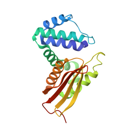
Mn(III)-PPIX bound Tt H-NOX


Experimental Data Snapshot

  • Method: X-RAY DIFFRACTION
  • Resolution: 2.10 Å
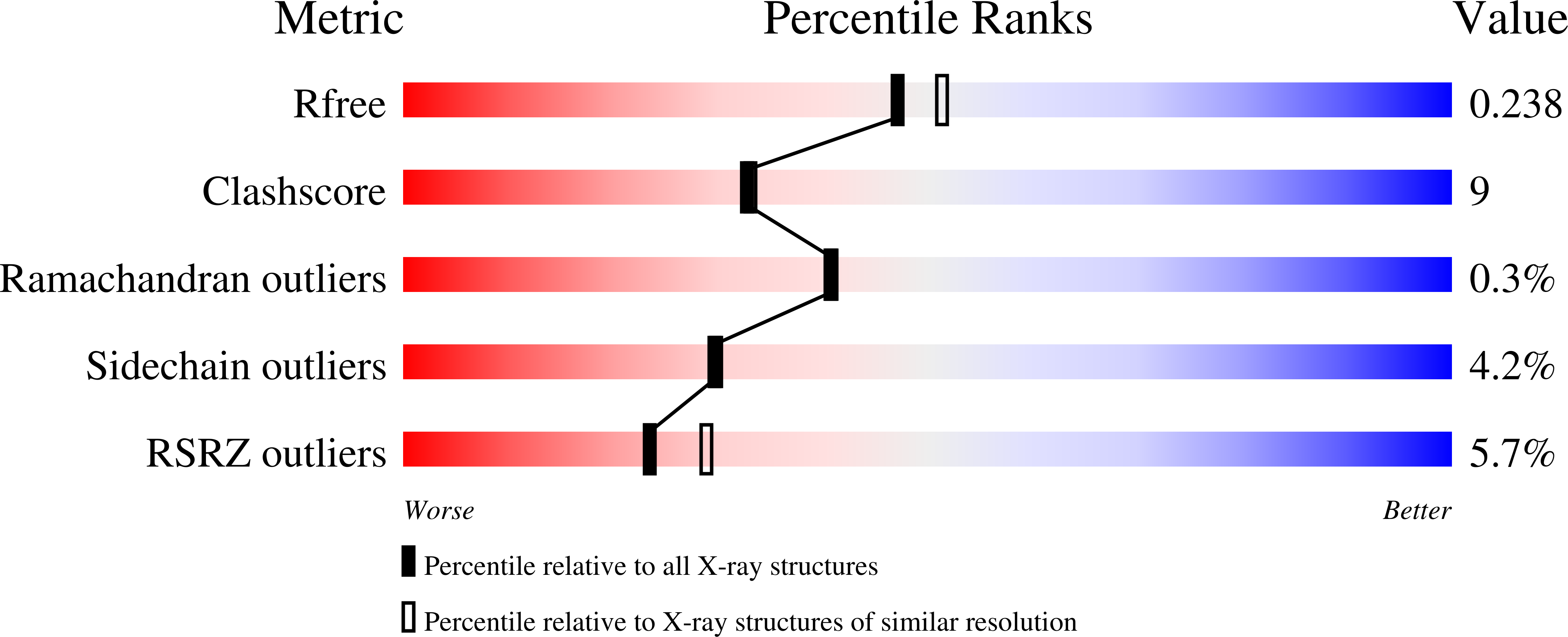
  • R-Value Free: 0.245 
  • R-Value Work: 0.209 
  • R-Value Observed: 0.211 

Starting Model: experimental
View more details

wwPDB Validation   3D Report Full Report


Ligand Structure Quality Assessment 


This is version 1.2 of the entry. See complete history


Literature

Porphyrin-Substituted H-NOX Proteins as High-Relaxivity MRI Contrast Agents.

Winter, M.B.Klemm, P.J.Phillips-Piro, C.M.Raymond, K.N.Marletta, M.A.

(2013) Inorg Chem 52: 2277-2279

  • DOI: https://doi.org/10.1021/ic302685h
  • Primary Citation of Related Structures:  
    4IT2

  • PubMed Abstract: 

    Heme proteins are exquisitely tuned to carry out diverse biological functions while employing identical heme cofactors. Although heme protein properties are often altered through modification of the protein scaffold, protein function can be greatly expanded and diversified through replacement of the native heme with an unnatural porphyrin of interest. Thus, porphyrin substitution in proteins affords new opportunities to rationally tailor heme protein chemical properties for new biological applications. Here, a highly thermally stable Heme Nitric oxide/OXygen binding (H-NOX) protein is evaluated as a magnetic resonance imaging (MRI) contrast agent. T1 and T2 relaxivities measured for the H-NOX protein containing its native heme are compared to the protein substituted with unnatural manganese(II/III) and gadolinium(III) porphyrins. H-NOX proteins are found to provide unique porphyrin coordination environments and have enhanced relaxivities compared to commercial small-molecule agents. Porphyrin substitution is a promising strategy to encapsulate MRI-active metals in heme protein scaffolds for future imaging applications.


  • Organizational Affiliation

    Department of Chemistry, California Institute for Quantitative Biosciences, Lawrence Berkeley National Laboratory, University of California, Berkeley, California 94720-3220, United States.


Macromolecules
Find similar proteins by:  (by identity cutoff)  |  3D Structure
Entity ID: 1
MoleculeChains Sequence LengthOrganismDetailsImage
Methyl-accepting chemotaxis protein
A, B
188Caldanaerobacter subterraneus subsp. tengcongensisMutation(s): 0 
Gene Names: Tar4TTE0680
UniProt
Find proteins for Q8RBX6 (Caldanaerobacter subterraneus subsp. tengcongensis (strain DSM 15242 / JCM 11007 / NBRC 100824 / MB4))
Explore Q8RBX6 
Go to UniProtKB:  Q8RBX6
Entity Groups  
Sequence Clusters30% Identity50% Identity70% Identity90% Identity95% Identity100% Identity
UniProt GroupQ8RBX6
Sequence Annotations
Expand
  • Reference Sequence
Experimental Data & Validation

Experimental Data

  • Method: X-RAY DIFFRACTION
  • Resolution: 2.10 Å
  • R-Value Free: 0.245 
  • R-Value Work: 0.209 
  • R-Value Observed: 0.211 
  • Space Group: C 1 2 1
Unit Cell:
Length ( Å )Angle ( ˚ )
a = 120.245α = 90
b = 45.416β = 122.82
c = 95.743γ = 90
Software Package:
Software NamePurpose
Blu-Icedata collection
PHASERphasing
PHENIXrefinement
HKL-2000data reduction
HKL-2000data scaling

Structure Validation

View Full Validation Report


Ligand Structure Quality Assessment 


Entry History 

Deposition Data

Revision History  (Full details and data files)

  • Version 1.0: 2013-02-27
    Type: Initial release
  • Version 1.1: 2013-03-20
    Changes: Database references
  • Version 1.2: 2023-09-20
    Changes: Data collection, Database references, Derived calculations, Refinement description