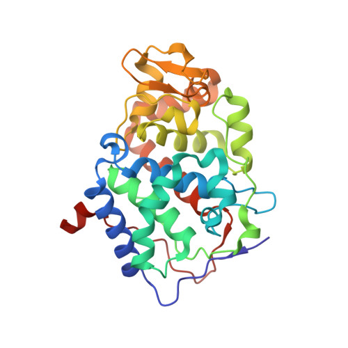
Blind prediction of charged ligand binding affinities in a model binding site.
Rocklin, G.J., Boyce, S.E., Fischer, M., Fish, I., Mobley, D.L., Shoichet, B.K., Dill, K.A.(2013) J Mol Biol 425: 4569-4583
- PubMed: 23896298
- DOI: https://doi.org/10.1016/j.jmb.2013.07.030
- Primary Citation of Related Structures:
4JM5, 4JM6, 4JM8, 4JM9, 4JMA, 4JMW, 4JPL, 4JPT, 4JPU, 4JQJ, 4JQK, 4JQM, 4JQN - PubMed Abstract:
Predicting absolute protein-ligand binding affinities remains a frontier challenge in ligand discovery and design. This becomes more difficult when ionic interactions are involved because of the large opposing solvation and electrostatic attraction energies. In a blind test, we examined whether alchemical free-energy calculations could predict binding affinities of 14 charged and 5 neutral compounds previously untested as ligands for a cavity binding site in cytochrome c peroxidase. In this simplified site, polar and cationic ligands compete with solvent to interact with a buried aspartate. Predictions were tested by calorimetry, spectroscopy, and crystallography. Of the 15 compounds predicted to bind, 13 were experimentally confirmed, while 4 compounds were false negative predictions. Predictions had a root-mean-square error of 1.95 kcal/mol to the experimental affinities, and predicted poses had an average RMSD of 1.7Å to the crystallographic poses. This test serves as a benchmark for these thermodynamically rigorous calculations at predicting binding affinities for charged compounds and gives insights into the existing sources of error, which are primarily electrostatic interactions inside proteins. Our experiments also provide a useful set of ionic binding affinities in a simplified system for testing new affinity prediction methods.
Organizational Affiliation:
Department of Pharmaceutical Chemistry, University of California San Francisco, 1700 4th Street, San Francisco, CA 94143-2550, USA; Biophysics Graduate Program, University of California San Francisco, 1700 4th Street, San Francisco, CA 94143-2550, USA.


