4JXC

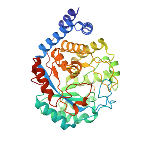
X-ray snapshots of possible intermediates in the time course of synthesis and degradation of protein-bound Fe4S4 clusters


Experimental Data Snapshot

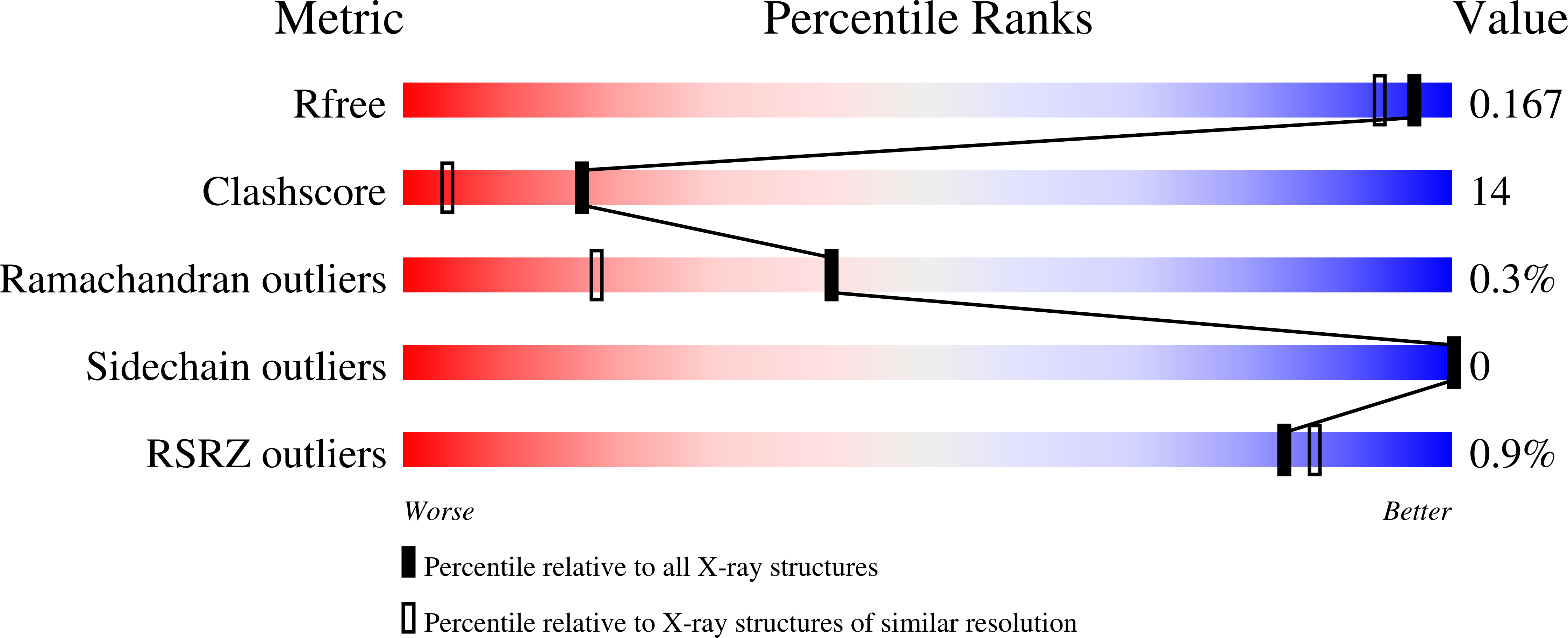
  • Method: X-RAY DIFFRACTION
  • Resolution: 1.50 Å
  • R-Value Free: 0.166 
  • R-Value Work: 0.121 
  • R-Value Observed: 0.124 

Starting Model: experimental
View more details

wwPDB Validation   3D Report Full Report


Ligand Structure Quality Assessment 


This is version 1.3 of the entry. See complete history


Literature

X-ray snapshots of possible intermediates in the time course of synthesis and degradation of protein-bound Fe4S4 clusters.

Nicolet, Y.Rohac, R.Martin, L.Fontecilla-Camps, J.C.

(2013) Proc Natl Acad Sci U S A 110: 7188-7192

  • DOI: https://doi.org/10.1073/pnas.1302388110
  • Primary Citation of Related Structures:  
    4JXC, 4JY8, 4JY9, 4JYD, 4JYE, 4JYF

  • PubMed Abstract: 

    Fe4S4 clusters are very common versatile prosthetic groups in proteins. Their redox property of being sensitive to O2-induced oxidative damage is, for instance, used by the cell to sense oxygen levels and switch between aerobic and anaerobic metabolisms, as exemplified by the fumarate, nitrate reduction regulator (FNR). Using the hydrogenase maturase HydE from Thermotoga maritima as a template, we obtained several unusual forms of FeS clusters, some of which are associated with important structural changes. These structures represent intermediate states relevant to both FeS cluster assembly and degradation. We observe one Fe2S2 cluster bound by two cysteine persulfide residues. This observation lends structural support to a very recent Raman study, which reported that Fe4S4-to-Fe2S2 cluster conversion upon oxygen exposure in FNR resulted in concomitant production of cysteine persulfide as cluster ligands. Similar persulfide ligands have been observed in vitro for several other Fe4S4 cluster-containing proteins. We have also monitored FeS cluster conversion directly in our protein crystals. Our structures indicate that the Fe4S4-to-Fe2S2 change requires large structural modifications, which are most likely responsible for the dimer-monomer transition in FNR.


  • Organizational Affiliation

    Metalloproteins Unit, Institut de Biologie Structurale J.-P. Ebel, Commissariat à l'Energie Atomique-Centre National de la Recherche Scientifique-Université Grenoble-Alpes, 38027 Grenoble, France. [email protected]


Macromolecules
Find similar proteins by:  (by identity cutoff)  |  3D Structure
Entity ID: 1
MoleculeChains Sequence LengthOrganismDetailsImage
FEFE-HYDROGENASE MATURASE348Thermotoga maritima MSB8Mutation(s): 0 
Gene Names: TM_1269ThemaDRAFT_0043
EC: 1.8
UniProt
Find proteins for Q9X0Z6 (Thermotoga maritima (strain ATCC 43589 / DSM 3109 / JCM 10099 / NBRC 100826 / MSB8))
Explore Q9X0Z6 
Go to UniProtKB:  Q9X0Z6
Entity Groups  
Sequence Clusters30% Identity50% Identity70% Identity90% Identity95% Identity100% Identity
UniProt GroupQ9X0Z6
Sequence Annotations
Expand
  • Reference Sequence
Small Molecules
Ligands 9 Unique
IDChains Name / Formula / InChI Key2D Diagram3D Interactions
1N7
Query on 1N7

Download Ideal Coordinates CCD File 
D [auth A],
E [auth A],
F [auth A],
G [auth A],
H [auth A]
CHAPSO
C32 H59 N2 O8 S
GUQQBLRVXOUDTN-XOHPMCGNSA-O
SAM
Query on SAM

Download Ideal Coordinates CCD File 
C [auth A]S-ADENOSYLMETHIONINE
C15 H22 N6 O5 S
MEFKEPWMEQBLKI-FCKMPRQPSA-N
SF4
Query on SF4

Download Ideal Coordinates CCD File 
B [auth A]IRON/SULFUR CLUSTER
Fe4 S4
LJBDFODJNLIPKO-UHFFFAOYSA-N
FES
Query on FES

Download Ideal Coordinates CCD File 
J [auth A]FE2/S2 (INORGANIC) CLUSTER
Fe2 S2
NIXDOXVAJZFRNF-UHFFFAOYSA-N
SO4
Query on SO4

Download Ideal Coordinates CCD File 
P [auth A]SULFATE ION
O4 S
QAOWNCQODCNURD-UHFFFAOYSA-L
GOL
Query on GOL

Download Ideal Coordinates CCD File 
I [auth A]GLYCEROL
C3 H8 O3
PEDCQBHIVMGVHV-UHFFFAOYSA-N
IPA
Query on IPA

Download Ideal Coordinates CCD File 
M [auth A]ISOPROPYL ALCOHOL
C3 H8 O
KFZMGEQAYNKOFK-UHFFFAOYSA-N
CL
Query on CL

Download Ideal Coordinates CCD File 
N [auth A],
O [auth A]
CHLORIDE ION
Cl
VEXZGXHMUGYJMC-UHFFFAOYSA-M
H2S
Query on H2S

Download Ideal Coordinates CCD File 
K [auth A],
L [auth A]
HYDROSULFURIC ACID
H2 S
RWSOTUBLDIXVET-UHFFFAOYSA-N
Modified Residues  1 Unique
IDChains TypeFormula2D DiagramParent
CSO
Query on CSO
A
L-PEPTIDE LINKINGC3 H7 N O3 SCYS
Experimental Data & Validation

Experimental Data

  • Method: X-RAY DIFFRACTION
  • Resolution: 1.50 Å
  • R-Value Free: 0.166 
  • R-Value Work: 0.121 
  • R-Value Observed: 0.124 
  • Space Group: P 21 21 21
Unit Cell:
Length ( Å )Angle ( ˚ )
a = 51.132α = 90
b = 78.729β = 90
c = 86.209γ = 90
Software Package:
Software NamePurpose
ADSCdata collection
PHASERphasing
REFMACrefinement
XDSdata reduction
XSCALEdata scaling

Structure Validation

View Full Validation Report


Ligand Structure Quality Assessment 


Entry History 

Deposition Data

Revision History  (Full details and data files)

  • Version 1.0: 2013-05-01
    Type: Initial release
  • Version 1.1: 2013-05-15
    Changes: Database references
  • Version 1.2: 2023-09-20
    Changes: Data collection, Database references, Derived calculations, Refinement description
  • Version 1.3: 2023-12-06
    Changes: Data collection