4KEY

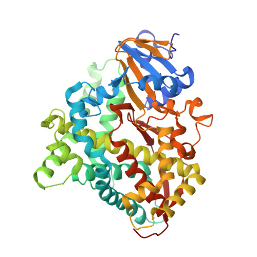
Structure of P450 BM3 A82F F87V in complex with omeprazole


Experimental Data Snapshot

  • Method: X-RAY DIFFRACTION
  • Resolution: 2.05 Å
  • R-Value Free: 0.235 
  • R-Value Work: 0.187 
  • R-Value Observed: 0.189 

wwPDB Validation   3D Report Full Report


Ligand Structure Quality Assessment 


This is version 1.2 of the entry. See complete history


Literature

Key Mutations Alter the Cytochrome P450 BM3 Conformational Landscape and Remove Inherent Substrate Bias.

Butler, C.F.Peet, C.Mason, A.E.Voice, M.W.Leys, D.Munro, A.W.

(2013) J Biol Chem 288: 25387-25399

  • DOI: https://doi.org/10.1074/jbc.M113.479717
  • Primary Citation of Related Structures:  
    4KEW, 4KEY, 4KF0, 4KF2

  • PubMed Abstract: 

    Cytochrome P450 monooxygenases (P450s) have enormous potential in the production of oxychemicals, due to their unparalleled regio- and stereoselectivity. The Bacillus megaterium P450 BM3 enzyme is a key model system, with several mutants (many distant from the active site) reported to alter substrate selectivity. It has the highest reported monooxygenase activity of the P450 enzymes, and this catalytic efficiency has inspired protein engineering to enable its exploitation for biotechnologically relevant oxidations with structurally diverse substrates. However, a structural rationale is lacking to explain how these mutations have such effects in the absence of direct change to the active site architecture. Here, we provide the first crystal structures of BM3 mutants in complex with a human drug substrate, the proton pump inhibitor omeprazole. Supported by solution data, these structures reveal how mutation alters the conformational landscape and decreases the free energy barrier for transition to the substrate-bound state. Our data point to the importance of such "gatekeeper" mutations in enabling major changes in substrate recognition. We further demonstrate that these mutants catalyze the same 5-hydroxylation reaction as performed by human CYP2C19, the major human omeprazole-metabolizing P450 enzyme.


  • Organizational Affiliation

    From the Manchester Institute of Biotechnology, Faculty of Life Sciences, University of Manchester, 131 Princess Street, Manchester M1 7DN, United Kingdom and.


Macromolecules
Find similar proteins by:  (by identity cutoff)  |  3D Structure
Entity ID: 1
MoleculeChains Sequence LengthOrganismDetailsImage
Bifunctional P-450/NADPH-P450 reductase
A, B
455Priestia megateriumMutation(s): 2 
Gene Names: cyp102A1cyp102
EC: 1.14.14.1 (PDB Primary Data), 1.6.2.4 (PDB Primary Data)
UniProt
Find proteins for P14779 (Priestia megaterium (strain ATCC 14581 / DSM 32 / CCUG 1817 / JCM 2506 / NBRC 15308 / NCIMB 9376 / NCTC 10342 / NRRL B-14308 / VKM B-512 / Ford 19))
Explore P14779 
Go to UniProtKB:  P14779
Entity Groups  
Sequence Clusters30% Identity50% Identity70% Identity90% Identity95% Identity100% Identity
UniProt GroupP14779
Sequence Annotations
Expand
  • Reference Sequence
Experimental Data & Validation

Experimental Data

  • Method: X-RAY DIFFRACTION
  • Resolution: 2.05 Å
  • R-Value Free: 0.235 
  • R-Value Work: 0.187 
  • R-Value Observed: 0.189 
  • Space Group: P 21 21 21
Unit Cell:
Length ( Å )Angle ( ˚ )
a = 59.31α = 90
b = 130.66β = 90
c = 145.98γ = 90
Software Package:
Software NamePurpose
ADSCdata collection
PHASERphasing
REFMACrefinement
XDSdata reduction
XDSdata scaling

Structure Validation

View Full Validation Report


Ligand Structure Quality Assessment 


Entry History 

Deposition Data

  • Released Date: 2013-07-10 
  • Deposition Author(s): Leys, D.

Revision History  (Full details and data files)

  • Version 1.0: 2013-07-10
    Type: Initial release
  • Version 1.1: 2013-10-02
    Changes: Database references
  • Version 1.2: 2024-02-28
    Changes: Data collection, Database references, Derived calculations, Structure summary