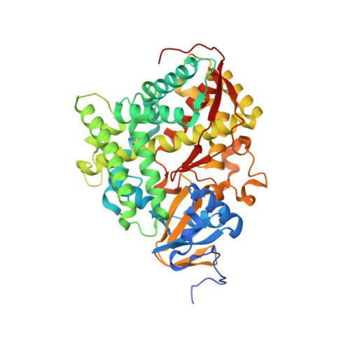
Structural Evidence: A Single Charged Residue Affects Substrate Binding in Cytochrome P450 BM-3.
Catalano, J., Sadre-Bazzaz, K., Amodeo, G.A., Tong, L., McDermott, A.(2013) Biochemistry 52: 6807-6815
- PubMed: 23829560
- DOI: https://doi.org/10.1021/bi4000645
- Primary Citation of Related Structures:
4KPA, 4KPB - PubMed Abstract:
Cytochrome P450 BM-3 is a bacterial enzyme with sequence similarity to mammalian P450s that catalyzes the hydroxylation of fatty acids with high efficiency. Enzyme-substrate binding and dynamics has been an important topic of study for cytochromes P450 because most of the crystal structures of substrate-bound structures show the complex in an inactive state. We have determined a new crystal structure for cytochrome P450 BM-3 in complex with N-palmitoylglycine (NPG), which unexpectedly showed a direct bidentate ion pair between NPG and arginine 47 (R47). We further explored the role of R47, the only charged residue in the binding pocket in cytochrome P450 BM-3, through mutagenesis and crystallographic studies. The mutations of R47 to glutamine (R47Q), glutamic acid (R47E), and lysine (R47K) were designed to investigate the role of its charge in binding and catalysis. The oppositely charged R47E mutation had the greatest effect on activity and binding. The crystal structure of R47E BMP shows that the glutamic acid side chain is blocking the entrance to the binding pocket, accounting for NPG's low binding affinity and charge repulsion. For R47Q and R47K BM-3, the mutations caused only a slight change in kcat and a large change in Km and Kd, which suggests that R47 mostly is involved in binding and that our crystal structure, 4KPA , represents an initial binding step in the P450 cycle.
Organizational Affiliation:
Department of Chemistry, Columbia University , 3000 Broadway, New York, New York 10027, United States.


