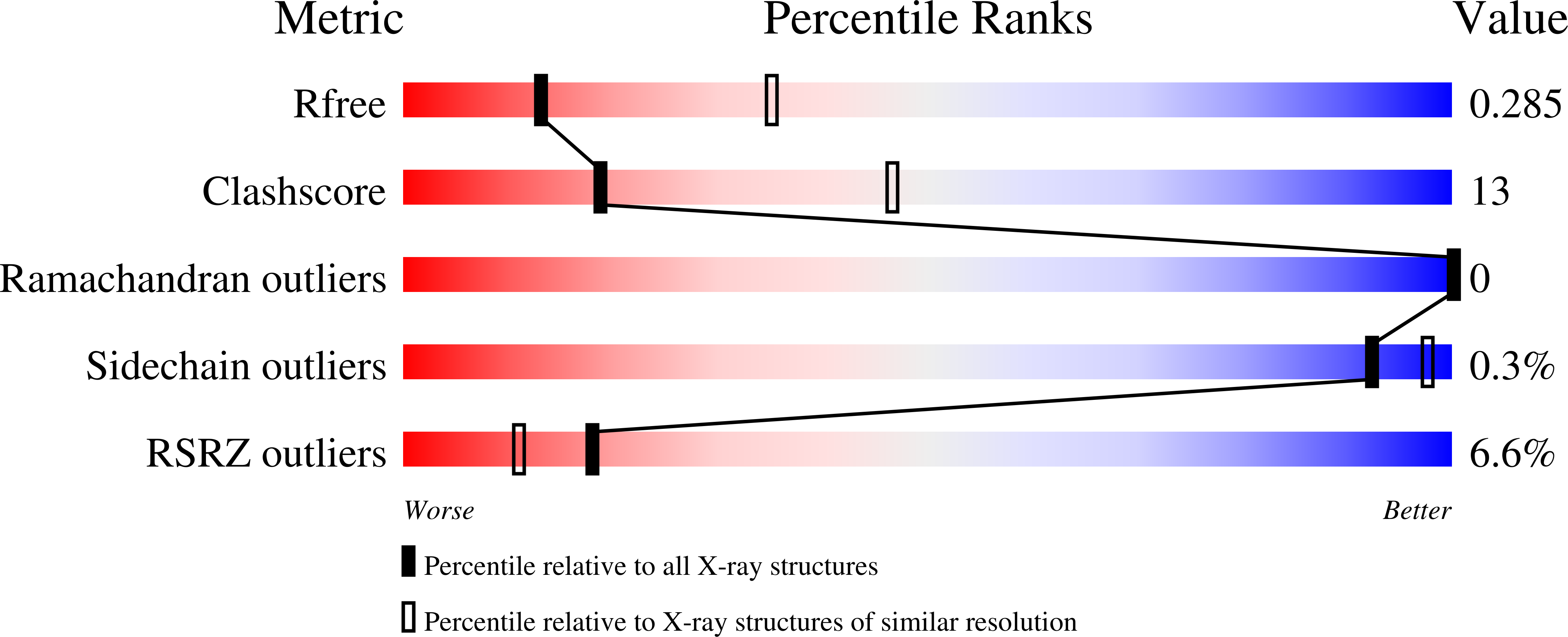
4M08

Crystal Structure of Mutant Chlorite Dismutase from Candidatus Nitrospira defluvii W145V


Experimental Data Snapshot

  • Method: X-RAY DIFFRACTION
  • Resolution: 2.80 Å
  • R-Value Free: 0.280 
  • R-Value Work: 0.221 
  • R-Value Observed: 0.224 

wwPDB Validation   3D Report Full Report


Ligand Structure Quality Assessment 


This is version 1.2 of the entry. See complete history


Literature

Manipulating conserved heme cavity residues of chlorite dismutase: effect on structure, redox chemistry, and reactivity.

Hofbauer, S.Gysel, K.Bellei, M.Hagmuller, A.Schaffner, I.Mlynek, G.Kostan, J.Pirker, K.F.Daims, H.Furtmuller, P.G.Battistuzzi, G.Djinovic-Carugo, K.Obinger, C.

(2014) Biochemistry 53: 77-89

  • DOI: https://doi.org/10.1021/bi401042z
  • Primary Citation of Related Structures:  
    4M05, 4M06, 4M07, 4M08, 4M09

  • PubMed Abstract: 

    Chlorite dismutases (Clds) are heme b containing oxidoreductases that convert chlorite to chloride and molecular oxygen. In order to elucidate the role of conserved heme cavity residues in the catalysis of this reaction comprehensive mutational and biochemical analyses of Cld from "Candidatus Nitrospira defluvii" (NdCld) were performed. Particularly, point mutations of the cavity-forming residues R173, K141, W145, W146, and E210 were performed. The effect of manipulation in 12 single and double mutants was probed by UV-vis spectroscopy, spectroelectrochemistry, pre-steady-state and steady-state kinetics, and X-ray crystallography. Resulting biochemical data are discussed with respect to the known crystal structure of wild-type NdCld and the variants R173A and R173K as well as the structures of R173E, W145V, W145F, and the R173Q/W146Y solved in this work. The findings allow a critical analysis of the role of these heme cavity residues in the reaction mechanism of chlorite degradation that is proposed to involve hypohalous acid as transient intermediate and formation of an O═O bond. The distal R173 is shown to be important (but not fully essential) for the reaction with chlorite, and, upon addition of cyanide, it acts as a proton acceptor in the formation of the resulting low-spin complex. The proximal H-bonding network including K141-E210-H160 keeps the enzyme in its ferric (E°' = -113 mV) and mainly five-coordinated high-spin state and is very susceptible to perturbation.


  • Organizational Affiliation

    Department of Chemistry, Division of Biochemistry, VIBT - Vienna Institute of BioTechnology, BOKU - University of Natural Resources and Life Sciences , A-1190 Vienna, Austria.


Macromolecules
Find similar proteins by:  (by identity cutoff)  |  3D Structure
Entity ID: 1
MoleculeChains Sequence LengthOrganismDetailsImage
Chlorite dismutase
A, B, C, D, E
241Nitrospira defluviiMutation(s): 1 
Gene Names: cldcld1NIDE1387
EC: 1.13.11.49
UniProt
Find proteins for B3U4H7 (Nitrospira defluvii)
Explore B3U4H7 
Go to UniProtKB:  B3U4H7
Entity Groups  
Sequence Clusters30% Identity50% Identity70% Identity90% Identity95% Identity100% Identity
UniProt GroupB3U4H7
Sequence Annotations
Expand
  • Reference Sequence
Small Molecules
Ligands 4 Unique
IDChains Name / Formula / InChI Key2D Diagram3D Interactions
HEM
Query on HEM

Download Ideal Coordinates CCD File 
AA [auth C],
F [auth A],
LA [auth D],
Q [auth B],
RA [auth E]
PROTOPORPHYRIN IX CONTAINING FE
C34 H32 Fe N4 O4
KABFMIBPWCXCRK-RGGAHWMASA-L
SO4
Query on SO4

Download Ideal Coordinates CCD File 
BA [auth C]
CA [auth C]
EA [auth C]
FA [auth C]
G [auth A]
BA [auth C],
CA [auth C],
EA [auth C],
FA [auth C],
G [auth A],
GA [auth C],
H [auth A],
HA [auth C],
I [auth A],
J [auth A],
K [auth A],
KA [auth D],
MA [auth D],
N [auth A],
NA [auth D],
O [auth A],
R [auth B],
S [auth B],
SA [auth E],
T [auth B],
TA [auth E],
Z [auth B]
SULFATE ION
O4 S
QAOWNCQODCNURD-UHFFFAOYSA-L
IMD
Query on IMD

Download Ideal Coordinates CCD File 
DA [auth C],
L [auth A],
OA [auth D],
UA [auth E],
W [auth B]
IMIDAZOLE
C3 H5 N2
RAXXELZNTBOGNW-UHFFFAOYSA-O
EDO
Query on EDO

Download Ideal Coordinates CCD File 
IA [auth C]
JA [auth C]
M [auth A]
P [auth A]
PA [auth E]
IA [auth C],
JA [auth C],
M [auth A],
P [auth A],
PA [auth E],
QA [auth E],
U [auth B],
V [auth B],
X [auth B],
Y [auth B]
1,2-ETHANEDIOL
C2 H6 O2
LYCAIKOWRPUZTN-UHFFFAOYSA-N
Experimental Data & Validation

Experimental Data

  • Method: X-RAY DIFFRACTION
  • Resolution: 2.80 Å
  • R-Value Free: 0.280 
  • R-Value Work: 0.221 
  • R-Value Observed: 0.224 
  • Space Group: P 32 2 1
Unit Cell:
Length ( Å )Angle ( ˚ )
a = 145.83α = 90
b = 145.83β = 90
c = 137.225γ = 120
Software Package:
Software NamePurpose
PHENIXrefinement
PDB_EXTRACTdata extraction
PROTEUM PLUSdata collection
PROTEUM2data reduction
SCALAdata scaling
MOLREPphasing

Structure Validation

View Full Validation Report


Ligand Structure Quality Assessment 


Entry History 

Deposition Data

Revision History  (Full details and data files)

  • Version 1.0: 2014-01-15
    Type: Initial release
  • Version 1.1: 2014-01-29
    Changes: Database references
  • Version 1.2: 2024-02-28
    Changes: Data collection, Database references, Derived calculations, Refinement description