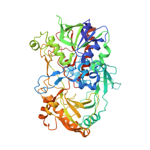
4MJW

Crystal Structure of Choline Oxidase in Complex with the Reaction Product Glycine Betaine


Experimental Data Snapshot

  • Method: X-RAY DIFFRACTION
  • Resolution: 1.95 Å
  • R-Value Free: 0.181 
  • R-Value Work: 0.151 
  • R-Value Observed: 0.152 

Starting Model: experimental
View more details

wwPDB Validation   3D Report Full Report


Ligand Structure Quality Assessment 


This is version 2.0 of the entry. See complete history


Literature

Structure of choline oxidase in complex with the reaction product glycine betaine.

Salvi, F.Wang, Y.F.Weber, I.T.Gadda, G.

(2014) Acta Crystallogr D Biol Crystallogr 70: 405-413

  • DOI: https://doi.org/10.1107/S1399004713029283
  • Primary Citation of Related Structures:  
    4MJW

  • PubMed Abstract: 

    Choline oxidase from Arthrobacter globiformis, which is involved in the biosynthesis of glycine betaine from choline, has been extensively characterized in its mechanistic and structural properties. Despite the knowledge gained on the enzyme, the details of substrate access to the active site are not fully understood. The `loop-and-lid' mechanism described for the glucose-methanol-choline enzyme superfamily has not been confirmed for choline oxidase. Instead, a hydrophobic cluster on the solvent-accessible surface of the enzyme has been proposed by molecular dynamics to control substrate access to the active site. Here, the crystal structure of the enzyme was solved in complex with glycine betaine at pH 6.0 at 1.95 Å resolution, allowing a structural description of the ligand-enzyme interactions in the active site. This structure is the first of choline oxidase in complex with a physiologically relevant ligand. The protein structures with and without ligand are virtually identical, with the exception of a loop at the dimer interface, which assumes two distinct conformations. The different conformations of loop 250-255 define different accessibilities of the proposed active-site entrance delimited by the hydrophobic cluster on the other subunit of the dimer, suggesting a role in regulating substrate access to the active site.


  • Organizational Affiliation

    Department of Chemistry, Georgia State University, PO Box 3965, Atlanta, GA 30302-3965, USA.


Macromolecules
Find similar proteins by:  (by identity cutoff)  |  3D Structure
Entity ID: 1
MoleculeChains Sequence LengthOrganismDetailsImage
Choline oxidase
A, B
546Arthrobacter globiformisMutation(s): 0 
Gene Names: codA
EC: 1.1.3.17
UniProt
Find proteins for Q7X2H8 (Arthrobacter globiformis)
Explore Q7X2H8 
Go to UniProtKB:  Q7X2H8
Entity Groups  
Sequence Clusters30% Identity50% Identity70% Identity90% Identity95% Identity100% Identity
UniProt GroupQ7X2H8
Sequence Annotations
Expand
  • Reference Sequence
Small Molecules
Ligands 4 Unique
IDChains Name / Formula / InChI Key2D Diagram3D Interactions
FAD
Query on FAD

Download Ideal Coordinates CCD File 
C [auth A],
H [auth B]
FLAVIN-ADENINE DINUCLEOTIDE
C27 H33 N9 O15 P2
VWWQXMAJTJZDQX-UYBVJOGSSA-N
BET
Query on BET

Download Ideal Coordinates CCD File 
D [auth A],
I [auth B]
TRIMETHYL GLYCINE
C5 H12 N O2
KWIUHFFTVRNATP-UHFFFAOYSA-O
GOL
Query on GOL

Download Ideal Coordinates CCD File 
F [auth A],
G [auth A],
K [auth B],
L [auth B]
GLYCEROL
C3 H8 O3
PEDCQBHIVMGVHV-UHFFFAOYSA-N
ACT
Query on ACT

Download Ideal Coordinates CCD File 
E [auth A],
J [auth B]
ACETATE ION
C2 H3 O2
QTBSBXVTEAMEQO-UHFFFAOYSA-M
Experimental Data & Validation

Experimental Data

  • Method: X-RAY DIFFRACTION
  • Resolution: 1.95 Å
  • R-Value Free: 0.181 
  • R-Value Work: 0.151 
  • R-Value Observed: 0.152 
  • Space Group: P 43 21 2
Unit Cell:
Length ( Å )Angle ( ˚ )
a = 87.35α = 90
b = 87.35β = 90
c = 353.484γ = 90
Software Package:
Software NamePurpose
HKL-2000data collection
PHASESphasing
REFMACrefinement
HKL-2000data reduction
HKL-2000data scaling

Structure Validation

View Full Validation Report


Ligand Structure Quality Assessment 


Entry History 

Deposition Data

Revision History  (Full details and data files)

  • Version 1.0: 2014-02-12
    Type: Initial release
  • Version 1.1: 2014-09-24
    Changes: Database references
  • Version 1.2: 2023-09-20
    Changes: Data collection, Database references, Derived calculations, Refinement description
  • Version 2.0: 2023-11-15
    Changes: Atomic model, Data collection