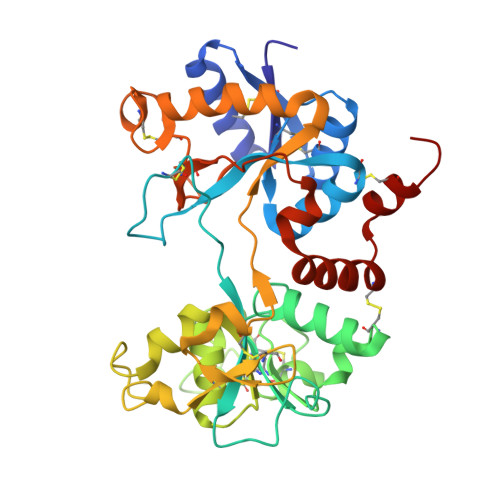
4OQO

Crystal structure of the tryptic generated iron-free C-lobe of bovine Lactoferrin at 2.42 Angstrom resolution


Experimental Data Snapshot

  • Method: X-RAY DIFFRACTION
  • Resolution: 2.42 Å
  • R-Value Free: 0.260 
  • R-Value Work: 0.223 
  • R-Value Observed: 0.225 

Starting Model: experimental
View more details

wwPDB Validation   3D Report Full Report


Ligand Structure Quality Assessment 


This is version 2.2 of the entry. See complete history


Literature

Structure of the iron-free true C-terminal half of bovine lactoferrin produced by tryptic digestion and its functional significance in the gut.

Rastogi, N.Singh, A.Pandey, S.N.Sinha, M.Bhushan, A.Kaur, P.Sharma, S.Singh, T.P.

(2014) FEBS J 281: 2871-2882

  • DOI: https://doi.org/10.1111/febs.12827
  • Primary Citation of Related Structures:  
    4OQO

  • PubMed Abstract: 

    Bovine lactoferrin, a 76-kDa glycoprotein (Ala1-Arg689) consists of two similar N- and C-terminal molecular halves with the ability to bind two Fe(3+) ions. The N-terminal half, designated as the N-lobe (Ala1-Arg341) and the C-terminal half designated as the C-lobe (Tyr342-Arg689) have similar iron-binding properties, but the resistant C-lobe prolongs the physiological role of bovine lactoferrin in the digestive tract. Here, we report the crystal structure of true C-lobe, which was produced by limited proteolysis of bovine lactoferrin using trypsin. In the first proteolysis step, two fragments of 21 kDa (Glu86-Lys282) and 45 kDa (Ser283-Arg689) were generated because two lysine residues, Lys85 and Lys282, in the structure of iron-saturated bovine lactoferrin were fully exposed. The 45-kDa fragment was further digested at the newly exposed side chain of Arg341, generating a 38-kDa perfect C-lobe (Tyr342-Arg689). By contrast, the apo-lactoferrin was cut by trypsin only at Arg341, which was exposed in the structure of apo-lactoferrin, whereas the other two sites with Lys85 and Lys282 are inaccessible. The purified iron-saturated C-lobe was crystallized at pH 4.0. The structure was determined by the molecular replacement method using coordinates of the C-terminal half (Arg342-Arg689) of intact camel apo-lactoferrin. The structure determination revealed that the iron atom was absent and the iron-binding cleft was found in a wide-open conformation, whereas in the previously determined structure of iron-saturated C-lobe of bovine lactoferrin, the iron atom was present and the iron-binding site was in the closed confirmation.


  • Organizational Affiliation

    Department of Biophysics, All India Institute of Medical Sciences, New Delhi, India.


Macromolecules
Find similar proteins by:  (by identity cutoff)  |  3D Structure
Entity ID: 1
MoleculeChains Sequence LengthOrganismDetailsImage
Lactotransferrin
A, B
348Bos taurusMutation(s): 0 
EC: 3.4.21
UniProt
Find proteins for P24627 (Bos taurus)
Explore P24627 
Go to UniProtKB:  P24627
Entity Groups  
Sequence Clusters30% Identity50% Identity70% Identity90% Identity95% Identity100% Identity
UniProt GroupP24627
Glycosylation
Glycosylation Sites: 2
Sequence Annotations
Expand
  • Reference Sequence
Oligosaccharides

Help

Entity ID: 2
MoleculeChains Length2D Diagram Glycosylation3D Interactions
2-acetamido-2-deoxy-beta-D-glucopyranose-(1-4)-2-acetamido-2-deoxy-beta-D-glucopyranose
C
2N-Glycosylation
Glycosylation Resources
GlyTouCan:  G42666HT
GlyCosmos:  G42666HT
GlyGen:  G42666HT
Experimental Data & Validation

Experimental Data

  • Method: X-RAY DIFFRACTION
  • Resolution: 2.42 Å
  • R-Value Free: 0.260 
  • R-Value Work: 0.223 
  • R-Value Observed: 0.225 
  • Space Group: P 1 21 1
Unit Cell:
Length ( Å )Angle ( ˚ )
a = 75.81α = 90
b = 49.44β = 94.33
c = 97.86γ = 90
Software Package:
Software NamePurpose
HKL-2000data collection
AMoREphasing
REFMACrefinement
HKL-2000data reduction
SCALEPACKdata scaling

Structure Validation

View Full Validation Report


Ligand Structure Quality Assessment 


Entry History 

Deposition Data

Revision History  (Full details and data files)

  • Version 1.0: 2014-03-12
    Type: Initial release
  • Version 1.1: 2014-07-16
    Changes: Database references
  • Version 2.0: 2020-07-29
    Type: Remediation
    Reason: Carbohydrate remediation
    Changes: Atomic model, Data collection, Derived calculations, Structure summary
  • Version 2.1: 2023-11-08
    Changes: Data collection, Database references, Refinement description, Structure summary
  • Version 2.2: 2024-10-30
    Changes: Structure summary