4TLF

Crystal structure of Thiol dioxygenase from Pseudomonas aeruginosa


Experimental Data Snapshot

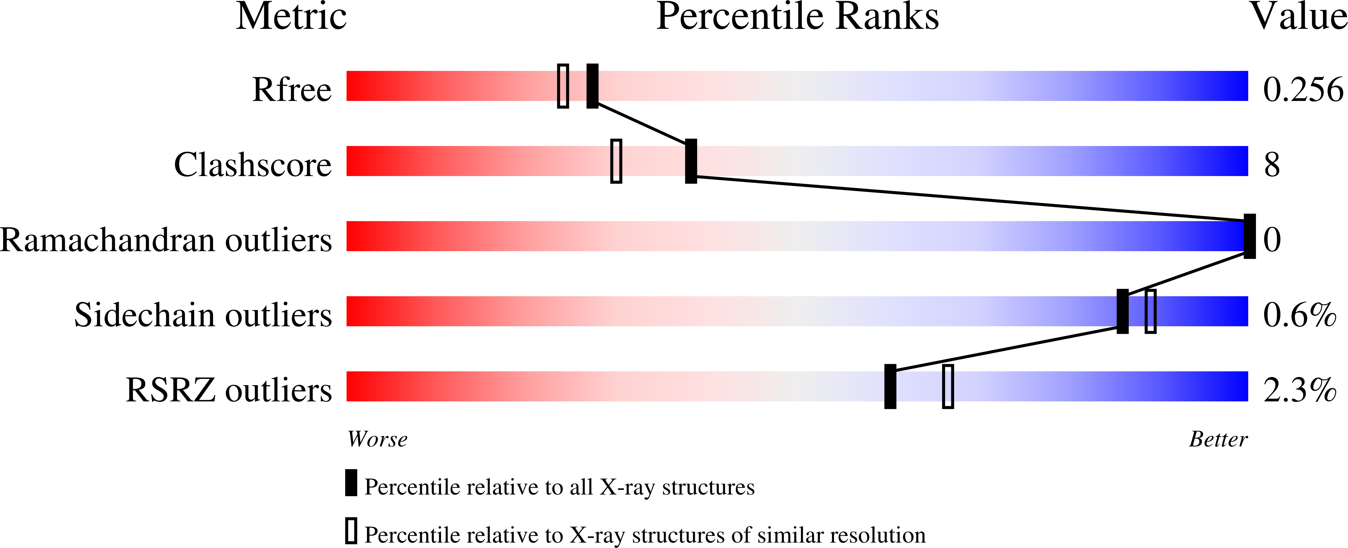
  • Method: X-RAY DIFFRACTION
  • Resolution: 2.14 Å
  • R-Value Free: 0.251 
  • R-Value Work: 0.198 
  • R-Value Observed: 0.201 

Starting Model: experimental
View more details

wwPDB Validation   3D Report Full Report


This is version 1.4 of the entry. See complete history


Literature

The Cysteine Dioxygenase Homologue from Pseudomonas aeruginosa Is a 3-Mercaptopropionate Dioxygenase.

Tchesnokov, E.P.Fellner, M.Siakkou, E.Kleffmann, T.Martin, L.W.Aloi, S.Lamont, I.L.Wilbanks, S.M.Jameson, G.N.

(2015) J Biol Chem 290: 24424-24437

  • DOI: https://doi.org/10.1074/jbc.M114.635672
  • Primary Citation of Related Structures:  
    4TLF

  • PubMed Abstract: 

    Thiol dioxygenation is the initial oxidation step that commits a thiol to important catabolic or biosynthetic pathways. The reaction is catalyzed by a family of specific non-heme mononuclear iron proteins each of which is reported to react efficiently with only one substrate. This family of enzymes includes cysteine dioxygenase, cysteamine dioxygenase, mercaptosuccinate dioxygenase, and 3-mercaptopropionate dioxygenase. Using sequence alignment to infer cysteine dioxygenase activity, a cysteine dioxygenase homologue from Pseudomonas aeruginosa (p3MDO) has been identified. Mass spectrometry of P. aeruginosa under standard growth conditions showed that p3MDO is expressed in low levels, suggesting that this metabolic pathway is available to the organism. Purified recombinant p3MDO is able to oxidize both cysteine and 3-mercaptopropionic acid in vitro, with a marked preference for 3-mercaptopropionic acid. We therefore describe this enzyme as a 3-mercaptopropionate dioxygenase. Mössbauer spectroscopy suggests that substrate binding to the ferrous iron is through the thiol but indicates that each substrate could adopt different coordination geometries. Crystallographic comparison with mammalian cysteine dioxygenase shows that the overall active site geometry is conserved but suggests that the different substrate specificity can be related to replacement of an arginine by a glutamine in the active site.


  • Organizational Affiliation

    From the Departments of Chemistry and.


Macromolecules
Find similar proteins by:  (by identity cutoff)  |  3D Structure
Entity ID: 1
MoleculeChains Sequence LengthOrganismDetailsImage
3-mercaptopropionate dioxygenase
A, B, C, D
211Pseudomonas aeruginosa PAO1Mutation(s): 0 
Gene Names: PA2602NC002516.2
EC: 1.13.11 (PDB Primary Data), 1.13.11.91 (UniProt)
UniProt
Find proteins for Q9I0N5 (Pseudomonas aeruginosa (strain ATCC 15692 / DSM 22644 / CIP 104116 / JCM 14847 / LMG 12228 / 1C / PRS 101 / PAO1))
Explore Q9I0N5 
Go to UniProtKB:  Q9I0N5
Entity Groups  
Sequence Clusters30% Identity50% Identity70% Identity90% Identity95% Identity100% Identity
UniProt GroupQ9I0N5
Sequence Annotations
Expand
  • Reference Sequence
Experimental Data & Validation

Experimental Data

  • Method: X-RAY DIFFRACTION
  • Resolution: 2.14 Å
  • R-Value Free: 0.251 
  • R-Value Work: 0.198 
  • R-Value Observed: 0.201 
  • Space Group: P 41 21 2
Unit Cell:
Length ( Å )Angle ( ˚ )
a = 66.942α = 90
b = 66.942β = 90
c = 377.354γ = 90
Software Package:
Software NamePurpose
PHENIXrefinement
XDSdata reduction
PDB_EXTRACTdata extraction
SCALAdata scaling
PHASERphasing

Structure Validation

View Full Validation Report


Entry History 

Deposition Data

Revision History  (Full details and data files)

  • Version 1.0: 2015-06-17
    Type: Initial release
  • Version 1.1: 2015-09-02
    Changes: Database references
  • Version 1.2: 2015-10-14
    Changes: Database references
  • Version 1.3: 2020-12-30
    Changes: Data collection, Derived calculations, Structure summary
  • Version 1.4: 2023-09-27
    Changes: Data collection, Database references, Refinement description