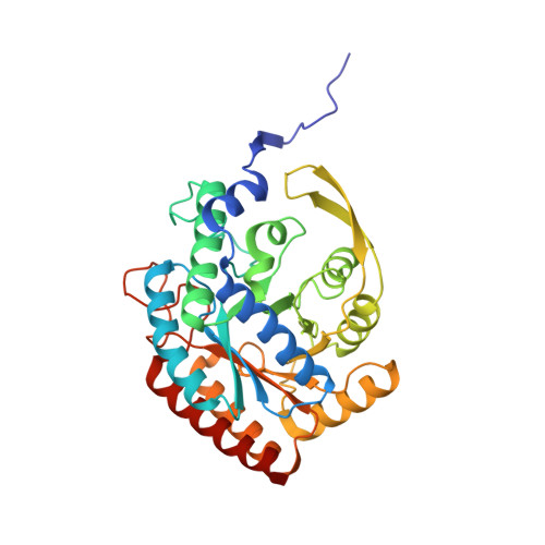
4UC5

Neisseria Meningitidis DAH7PS-Phenylalanine regulated


Experimental Data Snapshot

  • Method: X-RAY DIFFRACTION
  • Resolution: 2.19 Å
  • R-Value Free: 0.236 
  • R-Value Work: 0.189 
  • R-Value Observed: 0.191 

Starting Model: experimental
View more details

wwPDB Validation   3D Report Full Report


Ligand Structure Quality Assessment 


This is version 1.4 of the entry. See complete history


Literature

Calculated Pka Variations Expose Dynamic Allosteric Communication Networks.

Lang, E.J.Heyes, L.C.Jameson, G.B.Parker, E.J.

(2016) J Am Chem Soc 138: 2036

  • DOI: https://doi.org/10.1021/jacs.5b13134
  • Primary Citation of Related Structures:  
    4UC5

  • PubMed Abstract: 

    Allosteric regulation of protein function, the process by which binding of an effector molecule provokes a functional response from a distal site, is critical for metabolic pathways. Yet, the way the allosteric signal is communicated remains elusive, especially in dynamic, entropically driven regulation mechanisms for which no major conformational changes are observed. To identify these dynamic allosteric communication networks, we have developed an approach that monitors the pKa variations of ionizable residues over the course of molecular dynamics simulations performed in the presence and absence of an allosteric regulator. As the pKa of ionizable residues depends on their environment, it represents a simple metric to monitor changes in several complex factors induced by binding an allosteric effector. These factors include Coulombic interactions, hydrogen bonding, and solvation, as well as backbone motions and side chain fluctuations. The predictions that can be made with this method concerning the roles of ionizable residues for allosteric communication can then be easily tested experimentally by changing the working pH of the protein or performing single point mutations. To demonstrate the method's validity, we have applied this approach to the subtle dynamic regulation mechanism observed for Neisseria meningitidis 3-deoxy-d-arabino-heptulosonate 7-phosphate synthase, the first enzyme of aromatic biosynthesis. We were able to identify key communication pathways linking the allosteric binding site to the active site of the enzyme and to validate these findings experimentally by reestablishing the catalytic activity of allosterically inhibited enzyme via modulation of the working pH, without compromising the binding affinity of the allosteric regulator.


  • Organizational Affiliation

    Institute of Fundamental Sciences, Massey University , PO Box 11-222, Palmerston North 4422, New Zealand.


Macromolecules
Find similar proteins by:  (by identity cutoff)  |  3D Structure
Entity ID: 1
MoleculeChains Sequence LengthOrganismDetailsImage
PHOSPHO-2-DEHYDRO-3-DEOXYHEPTONATE ALDOLASE
A, B, C, D
351Neisseria meningitidisMutation(s): 0 
EC: 2.5.1.54
UniProt
Find proteins for Q9K169 (Neisseria meningitidis serogroup B (strain ATCC BAA-335 / MC58))
Explore Q9K169 
Go to UniProtKB:  Q9K169
Entity Groups  
Sequence Clusters30% Identity50% Identity70% Identity90% Identity95% Identity100% Identity
UniProt GroupQ9K169
Sequence Annotations
Expand
  • Reference Sequence
Small Molecules
Ligands 4 Unique
IDChains Name / Formula / InChI Key2D Diagram3D Interactions
PHE
Query on PHE

Download Ideal Coordinates CCD File 
I [auth A],
L [auth B],
N [auth C],
Q [auth D]
PHENYLALANINE
C9 H11 N O2
COLNVLDHVKWLRT-QMMMGPOBSA-N
PEG
Query on PEG

Download Ideal Coordinates CCD File 
F [auth A]DI(HYDROXYETHYL)ETHER
C4 H10 O3
MTHSVFCYNBDYFN-UHFFFAOYSA-N
SO4
Query on SO4

Download Ideal Coordinates CCD File 
G [auth A],
H [auth A],
K [auth B],
P [auth D]
SULFATE ION
O4 S
QAOWNCQODCNURD-UHFFFAOYSA-L
MN
Query on MN

Download Ideal Coordinates CCD File 
E [auth A],
J [auth B],
M [auth C],
O [auth D]
MANGANESE (II) ION
Mn
WAEMQWOKJMHJLA-UHFFFAOYSA-N
Experimental Data & Validation

Experimental Data

  • Method: X-RAY DIFFRACTION
  • Resolution: 2.19 Å
  • R-Value Free: 0.236 
  • R-Value Work: 0.189 
  • R-Value Observed: 0.191 
  • Space Group: P 1 21 1
Unit Cell:
Length ( Å )Angle ( ˚ )
a = 73.607α = 90
b = 143.46β = 96.18
c = 75.179γ = 90
Software Package:
Software NamePurpose
REFMACrefinement
MOSFLMdata reduction
SCALAdata scaling
PHASERphasing

Structure Validation

View Full Validation Report


Ligand Structure Quality Assessment 


Entry History 

Deposition Data

Revision History  (Full details and data files)

  • Version 1.0: 2015-11-25
    Type: Initial release
  • Version 1.1: 2015-12-02
    Changes: Database references
  • Version 1.2: 2016-02-03
    Changes: Database references
  • Version 1.3: 2016-03-02
    Changes: Database references
  • Version 1.4: 2023-12-20
    Changes: Data collection, Database references, Derived calculations, Other, Refinement description