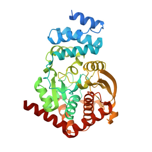
4X58

Anthranilate phosphoribosyl transferase variant N138A from Mycobacterium tuberculosis in complex with PRPP and Mg


Experimental Data Snapshot

  • Method: X-RAY DIFFRACTION
  • Resolution: 1.75 Å
  • R-Value Free: 0.222 
  • R-Value Work: 0.195 
  • R-Value Observed: 0.197 

Starting Model: experimental
View more details

wwPDB Validation   3D Report Full Report


Ligand Structure Quality Assessment 


This is version 1.2 of the entry. See complete history


Literature

Structures of Mycobacterium tuberculosis Anthranilate Phosphoribosyltransferase Variants Reveal the Conformational Changes That Facilitate Delivery of the Substrate to the Active Site.

Cookson, T.V.Evans, G.L.Castell, A.Baker, E.N.Lott, J.S.Parker, E.J.

(2015) Biochemistry 54: 6082-6092

  • DOI: https://doi.org/10.1021/acs.biochem.5b00612
  • Primary Citation of Related Structures:  
    4X58, 4X59, 4X5A, 4X5B, 4X5C, 4X5D, 4X5E

  • PubMed Abstract: 

    Anthranilate phosphoribosyltransferase (AnPRT) is essential for the biosynthesis of tryptophan in Mycobacterium tuberculosis (Mtb). This enzyme catalyzes the second committed step in tryptophan biosynthesis, the Mg²⁺-dependent reaction between 5'-phosphoribosyl-1'-pyrophosphate (PRPP) and anthranilate. The roles of residues predicted to be involved in anthranilate binding have been tested by the analysis of six Mtb-AnPRT variant proteins. Kinetic analysis showed that five of six variants were active and identified the conserved residue R193 as being crucial for both anthranilate binding and catalytic function. Crystal structures of these Mtb-AnPRT variants reveal the ability of anthranilate to bind in three sites along an extended anthranilate tunnel and expose the role of the mobile β2-α6 loop in facilitating the enzyme's sequential reaction mechanism. The β2-α6 loop moves sequentially between a "folded" conformation, partially occluding the anthranilate tunnel, via an "open" position to a "closed" conformation, which supports PRPP binding and allows anthranilate access via the tunnel to the active site. The return of the β2-α6 loop to the "folded" conformation completes the catalytic cycle, concordantly allowing the active site to eject the product PRA and rebind anthranilate at the opening of the anthranilate tunnel for subsequent reactions. Multiple anthranilate molecules blocking the anthranilate tunnel prevent the β2-α6 loop from undergoing the conformational changes required for catalysis, thus accounting for the unusual substrate inhibition of this enzyme.


  • Organizational Affiliation

    Maurice Wilkins Centre for Molecular Biodiscovery, Biomolecular Interaction Centre, and Department of Chemistry, University of Canterbury , 20 Kirkwood Avenue, Christchurch 8140, New Zealand.


Macromolecules
Find similar proteins by:  (by identity cutoff)  |  3D Structure
Entity ID: 1
MoleculeChains Sequence LengthOrganismDetailsImage
Anthranilate phosphoribosyltransferase
A, B
378Mycobacterium tuberculosisMutation(s): 1 
Gene Names: trpD
EC: 2.4.2.18
UniProt
Find proteins for P9WFX5 (Mycobacterium tuberculosis (strain ATCC 25618 / H37Rv))
Explore P9WFX5 
Go to UniProtKB:  P9WFX5
Entity Groups  
Sequence Clusters30% Identity50% Identity70% Identity90% Identity95% Identity100% Identity
UniProt GroupP9WFX5
Sequence Annotations
Expand
  • Reference Sequence
Small Molecules
Ligands 5 Unique
IDChains Name / Formula / InChI Key2D Diagram3D Interactions
PRP
Query on PRP

Download Ideal Coordinates CCD File 
G [auth A]1-O-pyrophosphono-5-O-phosphono-alpha-D-ribofuranose
C5 H13 O14 P3
PQGCEDQWHSBAJP-TXICZTDVSA-N
MLT
Query on MLT

Download Ideal Coordinates CCD File 
C [auth A]D-MALATE
C4 H6 O5
BJEPYKJPYRNKOW-UWTATZPHSA-N
GOL
Query on GOL

Download Ideal Coordinates CCD File 
D [auth A],
E [auth A],
F [auth A],
J [auth B]
GLYCEROL
C3 H8 O3
PEDCQBHIVMGVHV-UHFFFAOYSA-N
IMD
Query on IMD

Download Ideal Coordinates CCD File 
K [auth B]IMIDAZOLE
C3 H5 N2
RAXXELZNTBOGNW-UHFFFAOYSA-O
MG
Query on MG

Download Ideal Coordinates CCD File 
H [auth A],
I [auth A]
MAGNESIUM ION
Mg
JLVVSXFLKOJNIY-UHFFFAOYSA-N
Experimental Data & Validation

Experimental Data

  • Method: X-RAY DIFFRACTION
  • Resolution: 1.75 Å
  • R-Value Free: 0.222 
  • R-Value Work: 0.195 
  • R-Value Observed: 0.197 
  • Space Group: C 1 2 1
Unit Cell:
Length ( Å )Angle ( ˚ )
a = 94.739α = 90
b = 78.067β = 111.3
c = 101.331γ = 90
Software Package:
Software NamePurpose
REFMACrefinement
XDSdata reduction
PDB_EXTRACTdata extraction
Aimlessdata scaling
PHASERphasing

Structure Validation

View Full Validation Report


Ligand Structure Quality Assessment 


Entry History 

Deposition Data

Revision History  (Full details and data files)

  • Version 1.0: 2015-11-25
    Type: Initial release
  • Version 1.1: 2020-07-29
    Type: Remediation
    Reason: Carbohydrate remediation
    Changes: Data collection, Database references, Derived calculations, Structure summary
  • Version 1.2: 2023-09-27
    Changes: Data collection, Database references, Refinement description, Structure summary