4X6K

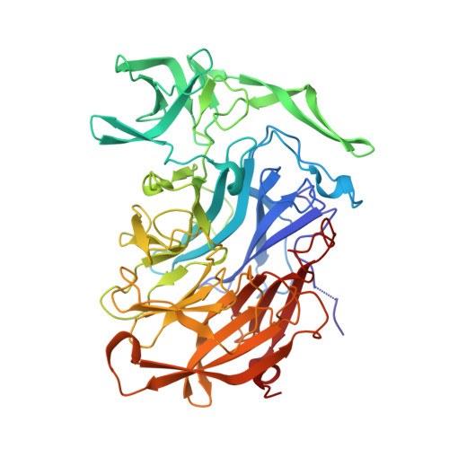
Crystal structure of the intramolecular trans-sialidase from Ruminococcus gnavus in complex with Siastatin B


Experimental Data Snapshot

  • Method: X-RAY DIFFRACTION
  • Resolution: 1.94 Å
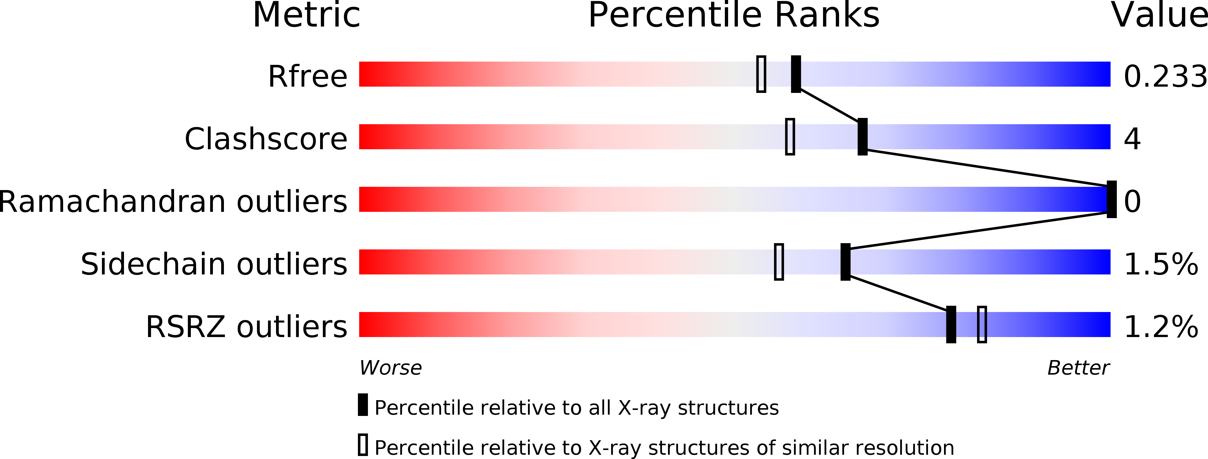
  • R-Value Free: 0.225 
  • R-Value Work: 0.177 
  • R-Value Observed: 0.179 

Starting Model: experimental
View more details

wwPDB Validation   3D Report Full Report


Ligand Structure Quality Assessment 


This is version 2.1 of the entry. See complete history


Literature

Discovery of intramolecular trans-sialidases in human gut microbiota suggests novel mechanisms of mucosal adaptation.

Tailford, L.E.Owen, C.D.Walshaw, J.Crost, E.H.Hardy-Goddard, J.Le Gall, G.de Vos, W.M.Taylor, G.L.Juge, N.

(2015) Nat Commun 6: 7624-7624

  • DOI: https://doi.org/10.1038/ncomms8624
  • Primary Citation of Related Structures:  
    4X47, 4X49, 4X4A, 4X6K

  • PubMed Abstract: 

    The gastrointestinal mucus layer is colonized by a dense community of microbes catabolizing dietary and host carbohydrates during their expansion in the gut. Alterations in mucosal carbohydrate availability impact on the composition of microbial species. Ruminococcus gnavus is a commensal anaerobe present in the gastrointestinal tract of >90% of humans and overrepresented in inflammatory bowel diseases (IBD). Using a combination of genomics, enzymology and crystallography, we show that the mucin-degrader R. gnavus ATCC 29149 strain produces an intramolecular trans-sialidase (IT-sialidase) that cleaves off terminal α2-3-linked sialic acid from glycoproteins, releasing 2,7-anhydro-Neu5Ac instead of sialic acid. Evidence of IT-sialidases in human metagenomes indicates that this enzyme occurs in healthy subjects but is more prevalent in IBD metagenomes. Our results uncover a previously unrecognized enzymatic activity in the gut microbiota, which may contribute to the adaptation of intestinal bacteria to the mucosal environment in health and disease.


  • Organizational Affiliation

    The Gut Health and Food Safety Institute Strategic Programme, Institute of Food Research, Norwich Research Park, Norwich NR4 7UA, UK.


Macromolecules
Find similar proteins by:  (by identity cutoff)  |  3D Structure
Entity ID: 1
MoleculeChains Sequence LengthOrganismDetailsImage
Anhydrosialidase486Mediterraneibacter gnavus CC55_001CMutation(s): 0 
Gene Names: HMPREF1201_01421
Entity Groups  
Sequence Clusters30% Identity50% Identity70% Identity90% Identity95% Identity100% Identity
Sequence Annotations
Expand
  • Reference Sequence
Small Molecules
Ligands 2 Unique
IDChains Name / Formula / InChI Key2D Diagram3D Interactions
3XR
Query on 3XR

Download Ideal Coordinates CCD File 
C [auth A](2S,3R,4S,5S)-2-(acetylamino)-5-carboxy-3,4-dihydroxypiperidinium
C8 H15 N2 O5
DQTKLICLJUKNCG-ZTYPAOSTSA-O
ACE
Query on ACE

Download Ideal Coordinates CCD File 
B [auth A]ACETYL GROUP
C2 H4 O
IKHGUXGNUITLKF-UHFFFAOYSA-N
Experimental Data & Validation

Experimental Data

  • Method: X-RAY DIFFRACTION
  • Resolution: 1.94 Å
  • R-Value Free: 0.225 
  • R-Value Work: 0.177 
  • R-Value Observed: 0.179 
  • Space Group: H 3
Unit Cell:
Length ( Å )Angle ( ˚ )
a = 99.109α = 90
b = 99.109β = 90
c = 131.708γ = 120
Software Package:
Software NamePurpose
REFMACrefinement
SCALEPACKdata scaling
PHASERphasing
PDB_EXTRACTdata extraction
Cootmodel building
HKL-2000data reduction

Structure Validation

View Full Validation Report


Ligand Structure Quality Assessment 


Entry History & Funding Information

Deposition Data


Funding OrganizationLocationGrant Number
Biotechnology and Biological Sciences Research CouncilUnited KingdomBB/J004529/1
Biotechnology and Biological Sciences Research CouncilUnited KingdomBB/L008602/1

Revision History  (Full details and data files)

  • Version 1.0: 2015-07-22
    Type: Initial release
  • Version 2.0: 2017-08-30
    Changes: Advisory, Atomic model, Author supporting evidence, Derived calculations
  • Version 2.1: 2024-01-10
    Changes: Data collection, Database references, Refinement description