4XC7

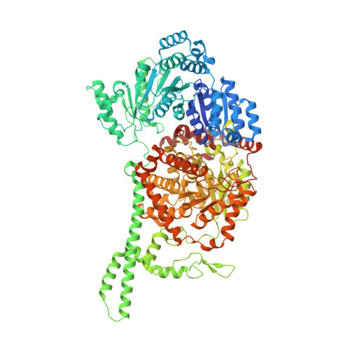
Isobutyryl-CoA mutase fused with bound butyryl-CoA and without cobalamin or GDP (apo-IcmF)


Experimental Data Snapshot

  • Method: X-RAY DIFFRACTION
  • Resolution: 3.45 Å
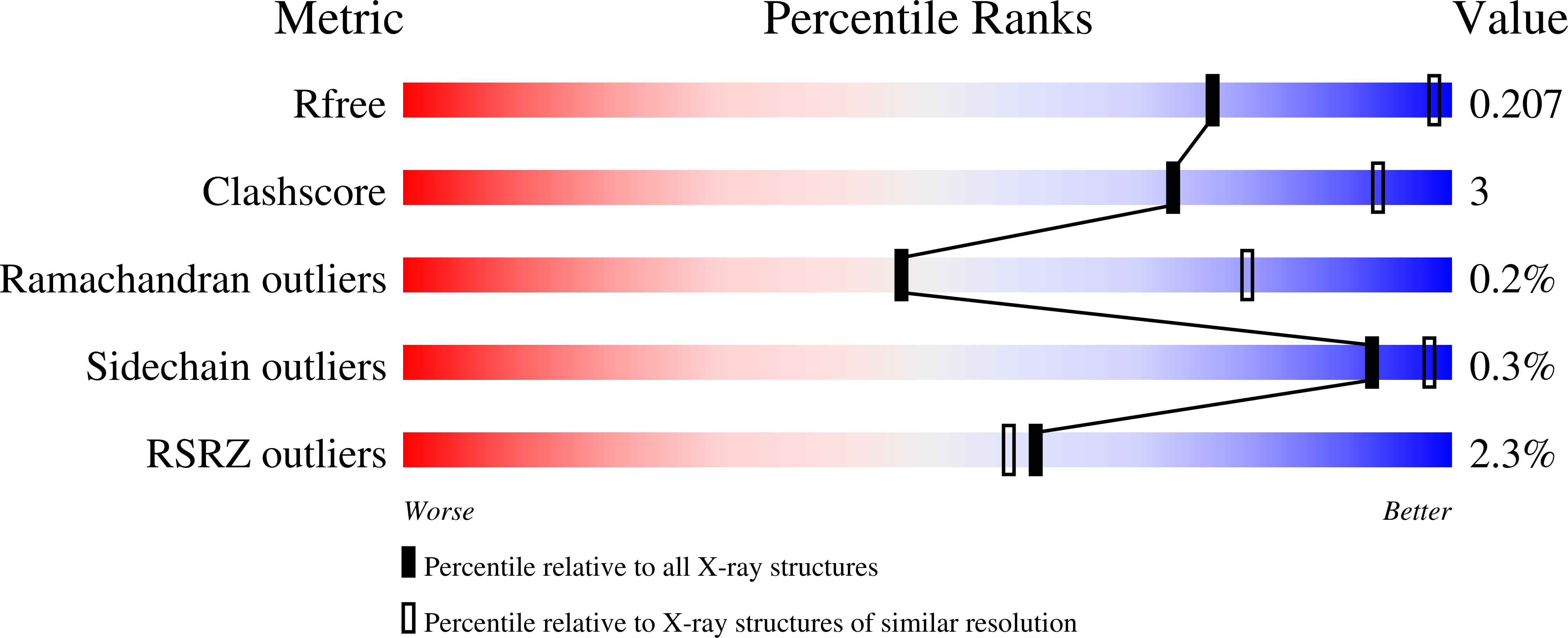
  • R-Value Free: 0.203 
  • R-Value Work: 0.186 
  • R-Value Observed: 0.187 

Starting Model: experimental
View more details

wwPDB Validation   3D Report Full Report


Ligand Structure Quality Assessment 


This is version 1.5 of the entry. See complete history


Literature

Visualization of a radical B12 enzyme with its G-protein chaperone.

Jost, M.Cracan, V.Hubbard, P.A.Banerjee, R.Drennan, C.L.

(2015) Proc Natl Acad Sci U S A 112: 2419-2424

  • DOI: https://doi.org/10.1073/pnas.1419582112
  • Primary Citation of Related Structures:  
    4XC6, 4XC7, 4XC8

  • PubMed Abstract: 

    G-protein metallochaperones ensure fidelity during cofactor assembly for a variety of metalloproteins, including adenosylcobalamin (AdoCbl)-dependent methylmalonyl-CoA mutase and hydrogenase, and thus have both medical and biofuel development applications. Here, we present crystal structures of IcmF, a natural fusion protein of AdoCbl-dependent isobutyryl-CoA mutase and its corresponding G-protein chaperone, which reveal the molecular architecture of a G-protein metallochaperone in complex with its target protein. These structures show that conserved G-protein elements become ordered upon target protein association, creating the molecular pathways that both sense and report on the cofactor loading state. Structures determined of both apo- and holo-forms of IcmF depict both open and closed enzyme states, in which the cofactor-binding domain is alternatively positioned for cofactor loading and for catalysis. Notably, the G protein moves as a unit with the cofactor-binding domain, providing a visualization of how a chaperone assists in the sequestering of a precious cofactor inside an enzyme active site.


  • Organizational Affiliation

    Department of Chemistry and.


Macromolecules
Find similar proteins by:  (by identity cutoff)  |  3D Structure
Entity ID: 1
MoleculeChains Sequence LengthOrganismDetailsImage
Isobutyryl-CoA mutase fused
A, B
1,113Cupriavidus metallidurans CH34Mutation(s): 0 
Gene Names: sbmRmet_0210
EC: 5.4.99.13 (PDB Primary Data), 3.6.5 (UniProt)
UniProt
Find proteins for Q1LRY0 (Cupriavidus metallidurans (strain ATCC 43123 / DSM 2839 / NBRC 102507 / CH34))
Explore Q1LRY0 
Go to UniProtKB:  Q1LRY0
Entity Groups  
Sequence Clusters30% Identity50% Identity70% Identity90% Identity95% Identity100% Identity
UniProt GroupQ1LRY0
Sequence Annotations
Expand
  • Reference Sequence
Experimental Data & Validation

Experimental Data

  • Method: X-RAY DIFFRACTION
  • Resolution: 3.45 Å
  • R-Value Free: 0.203 
  • R-Value Work: 0.186 
  • R-Value Observed: 0.187 
  • Space Group: H 3 2
Unit Cell:
Length ( Å )Angle ( ˚ )
a = 318.18α = 90
b = 318.18β = 90
c = 344.71γ = 120
Software Package:
Software NamePurpose
PHENIXrefinement
XDSdata reduction
XSCALEdata scaling
PHASERphasing
Cootmodel building

Structure Validation

View Full Validation Report


Ligand Structure Quality Assessment 


Entry History & Funding Information

Deposition Data


Funding OrganizationLocationGrant Number
National Institutes of Health/National Institute of General Medical Sciences (NIH/NIGMS)United StatesGM069857
Howard Hughes Medical Institute (HHMI)United States--

Revision History  (Full details and data files)

  • Version 1.0: 2015-02-11
    Type: Initial release
  • Version 1.1: 2015-03-04
    Changes: Database references
  • Version 1.2: 2016-01-27
    Changes: Non-polymer description
  • Version 1.3: 2017-09-13
    Changes: Author supporting evidence, Derived calculations
  • Version 1.4: 2019-11-20
    Changes: Author supporting evidence
  • Version 1.5: 2023-09-27
    Changes: Data collection, Database references, Refinement description