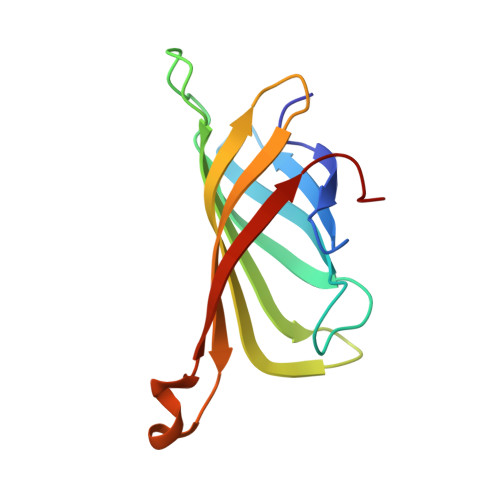
4Y5D

CRYSTAL STRUCTURE OF ALiS2-STREPTAVIDIN COMPLEX


Experimental Data Snapshot

  • Method: X-RAY DIFFRACTION
  • Resolution: 1.20 Å
  • R-Value Free: 0.174 
  • R-Value Work: 0.138 
  • R-Value Observed: 0.140 

wwPDB Validation   3D Report Full Report


Ligand Structure Quality Assessment 


This is version 1.3 of the entry. See complete history


Literature

Artificial Ligands of Streptavidin (ALiS): Discovery, Characterization, and Application for Reversible Control of Intracellular Protein Transport

Terai, T.Kohno, M.Boncompain, G.Sugiyama, S.Saito, N.Fujikake, R.Ueno, T.Komatsu, T.Hanaoka, K.Okabe, T.Urano, Y.Perez, F.Nagano, T.

(2015) J Am Chem Soc 137: 10464-10467

  • DOI: https://doi.org/10.1021/jacs.5b05672
  • Primary Citation of Related Structures:  
    4Y59, 4Y5D

  • PubMed Abstract: 

    Artificial ligands of streptavidin (ALiS) with association constants of ∼10(6) M(-1) were discovered by high-throughput screening of our chemical library, and their binding characteristics, including X-ray crystal structure of the streptavidin complex, were determined. Unlike biotin and its derivatives, ALiS exhibits fast dissociation kinetics and excellent cell permeability. The streptavidin-ALiS system provides a novel, practical compound-dependent methodology for repeated reversible cycling of protein localization between intracellular organella.


  • Organizational Affiliation

    Graduate School of Science, Osaka University , 2-1 Yamadaoka, Suita, Osaka 565-0871, Japan.


Macromolecules
Find similar proteins by:  (by identity cutoff)  |  3D Structure
Entity ID: 1
MoleculeChains Sequence LengthOrganismDetailsImage
Streptavidin
A, B, C, D
122Streptomyces avidiniiMutation(s): 0 
UniProt
Find proteins for P22629 (Streptomyces avidinii)
Explore P22629 
Go to UniProtKB:  P22629
Entity Groups  
Sequence Clusters30% Identity50% Identity70% Identity90% Identity95% Identity100% Identity
UniProt GroupP22629
Sequence Annotations
Expand
  • Reference Sequence
Small Molecules
Ligands 4 Unique
IDChains Name / Formula / InChI Key2D Diagram3D Interactions
PE3
Query on PE3

Download Ideal Coordinates CCD File 
F [auth A]3,6,9,12,15,18,21,24,27,30,33,36,39-TRIDECAOXAHENTETRACONTANE-1,41-DIOL
C28 H58 O15
ILLKMACMBHTSHP-UHFFFAOYSA-N
P6G
Query on P6G

Download Ideal Coordinates CCD File 
I [auth C],
J [auth C]
HEXAETHYLENE GLYCOL
C12 H26 O7
IIRDTKBZINWQAW-UHFFFAOYSA-N
MT6
Query on MT6

Download Ideal Coordinates CCD File 
E [auth A],
G [auth B],
K [auth D]
methyl 3-(4-oxo-4,5-dihydrofuro[3,2-c]pyridin-2-yl)benzoate
C15 H11 N O4
OVTGPKXYLOQTEQ-UHFFFAOYSA-N
DMS
Query on DMS

Download Ideal Coordinates CCD File 
H [auth C]DIMETHYL SULFOXIDE
C2 H6 O S
IAZDPXIOMUYVGZ-UHFFFAOYSA-N
Experimental Data & Validation

Experimental Data

  • Method: X-RAY DIFFRACTION
  • Resolution: 1.20 Å
  • R-Value Free: 0.174 
  • R-Value Work: 0.138 
  • R-Value Observed: 0.140 
  • Space Group: P 1 21 1
Unit Cell:
Length ( Å )Angle ( ˚ )
a = 57.927α = 90
b = 83.774β = 98.75
c = 45.947γ = 90
Software Package:
Software NamePurpose
REFMACrefinement
HKL-2000data reduction
HKL-2000data scaling
MOLREPphasing

Structure Validation

View Full Validation Report


Ligand Structure Quality Assessment 


Entry History 

Deposition Data

Revision History  (Full details and data files)

  • Version 1.0: 2015-09-23
    Type: Initial release
  • Version 1.1: 2015-12-16
    Changes: Database references
  • Version 1.2: 2020-02-05
    Changes: Data collection, Derived calculations
  • Version 1.3: 2024-03-20
    Changes: Data collection, Database references