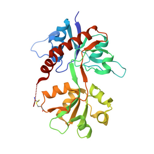
4YMB

Structure of the ligand-binding domain of GluK1 in complex with the antagonist CNG10111


Experimental Data Snapshot

  • Method: X-RAY DIFFRACTION
  • Resolution: 1.93 Å
  • R-Value Free: 0.220 
  • R-Value Work: 0.179 
  • R-Value Observed: 0.181 

Starting Model: experimental
View more details

wwPDB Validation   3D Report Full Report


Ligand Structure Quality Assessment 


This is version 1.4 of the entry. See complete history


Literature

Structure-Activity Relationship Study of Ionotropic Glutamate Receptor Antagonist (2S,3R)-3-(3-Carboxyphenyl)pyrrolidine-2-carboxylic Acid.

Krogsgaard-Larsen, N.Storgaard, M.Moller, C.Demmer, C.S.Hansen, J.Han, L.Monrad, R.N.Nielsen, B.Tapken, D.Pickering, D.S.Kastrup, J.S.Frydenvang, K.Bunch, L.

(2015) J Med Chem 58: 6131-6150

  • DOI: https://doi.org/10.1021/acs.jmedchem.5b00750
  • Primary Citation of Related Structures:  
    4YMA, 4YMB

  • PubMed Abstract: 

    Herein we describe the first structure-activity relationship study of the broad-range iGluR antagonist (2S,3R)-3-(3-carboxyphenyl)pyrrolidine-2-carboxylic acid (1) by exploring the pharmacological effect of substituents in the 4, 4', or 5' positions and the bioisosteric substitution of the distal carboxylic acid for a phosphonic acid moiety. Of particular interest is a hydroxyl group in the 4' position 2a which induced a preference in binding affinity for homomeric GluK3 over GluK1 (Ki = 0.87 and 4.8 μM, respectively). Two X-ray structures of ligand binding domains were obtained: 2e in GluA2-LBD and 2f in GluK1-LBD, both at 1.9 Å resolution. Compound 2e induces a D1-D2 domain opening in GluA2-LBD of 17.3-18.8° and 2f a domain opening in GluK1-LBD of 17.0-17.5° relative to the structures with glutamate. The pyrrolidine-2-carboxylate moiety of 2e and 2f shows a similar binding mode as kainate. The 3-carboxyphenyl ring of 2e and 2f forms contacts comparable to those of the distal carboxylate in kainate.


  • Organizational Affiliation

    †Chemical Neuroscience Group, ‡Biostructural Research Group, §Medicinal Chemistry Group, ∥Molecular, Cellular Pharmacology Group, Department of Drug Design and Pharmacology, Faculty of Health and Medical Sciences, University of Copenhagen, Universitetsparken 2, 2100 Copenhagen Ø, Denmark.


Macromolecules
Find similar proteins by:  (by identity cutoff)  |  3D Structure
Entity ID: 1
MoleculeChains Sequence LengthOrganismDetailsImage
Glutamate receptor ionotropic, kainate 1,Glutamate receptor ionotropic, kainate 1
A, B
257Rattus norvegicusMutation(s): 1 
Gene Names: Grik1Glur5
UniProt
Find proteins for P22756 (Rattus norvegicus)
Explore P22756 
Go to UniProtKB:  P22756
Entity Groups  
Sequence Clusters30% Identity50% Identity70% Identity90% Identity95% Identity100% Identity
UniProt GroupP22756
Sequence Annotations
Expand
  • Reference Sequence
Small Molecules
Ligands 5 Unique
IDChains Name / Formula / InChI Key2D Diagram3D Interactions
4E7
Query on 4E7

Download Ideal Coordinates CCD File 
C [auth A],
I [auth B]
(3R,4S)-3-(3-carboxyphenyl)-4-propyl-L-proline
C15 H19 N O4
OORLOXOWCACWSS-AGIUHOORSA-N
PEG
Query on PEG

Download Ideal Coordinates CCD File 
H [auth A]DI(HYDROXYETHYL)ETHER
C4 H10 O3
MTHSVFCYNBDYFN-UHFFFAOYSA-N
EDO
Query on EDO

Download Ideal Coordinates CCD File 
N [auth B]1,2-ETHANEDIOL
C2 H6 O2
LYCAIKOWRPUZTN-UHFFFAOYSA-N
ACT
Query on ACT

Download Ideal Coordinates CCD File 
D [auth A],
J [auth B],
K [auth B],
L [auth B]
ACETATE ION
C2 H3 O2
QTBSBXVTEAMEQO-UHFFFAOYSA-M
CL
Query on CL

Download Ideal Coordinates CCD File 
E [auth A],
F [auth A],
G [auth A],
M [auth B]
CHLORIDE ION
Cl
VEXZGXHMUGYJMC-UHFFFAOYSA-M
Experimental Data & Validation

Experimental Data

  • Method: X-RAY DIFFRACTION
  • Resolution: 1.93 Å
  • R-Value Free: 0.220 
  • R-Value Work: 0.179 
  • R-Value Observed: 0.181 
  • Space Group: P 41 21 2
Unit Cell:
Length ( Å )Angle ( ˚ )
a = 71.813α = 90
b = 71.813β = 90
c = 230.126γ = 90
Software Package:
Software NamePurpose
SCALAdata scaling
PHASERphasing
PHENIXrefinement
PDB_EXTRACTdata extraction
XDSdata reduction
Cootmodel building

Structure Validation

View Full Validation Report


Ligand Structure Quality Assessment 


Entry History & Funding Information

Deposition Data


Funding OrganizationLocationGrant Number
Denmark--

Revision History  (Full details and data files)

  • Version 1.0: 2015-08-05
    Type: Initial release
  • Version 1.1: 2015-08-26
    Changes: Database references
  • Version 1.2: 2017-08-09
    Changes: Data collection, Database references
  • Version 1.3: 2024-01-10
    Changes: Data collection, Database references, Refinement description
  • Version 1.4: 2024-11-06
    Changes: Structure summary