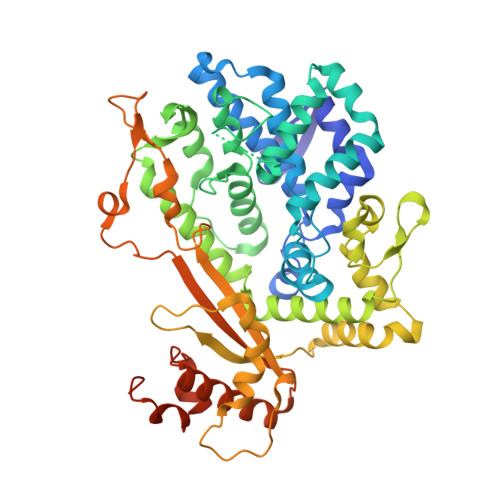
4ZWE

Crystal structure of the dGTP-bound catalytic core of SAMHD1 T592V mutant

  • Classification: HYDROLASE
  • Organism(s): Homo sapiens
  • Expression System: Escherichia coli
  • Mutation(s): Yes 

  • Deposited: 2015-05-19 Released: 2015-09-02 
  • Deposition Author(s): Tang, C., Ji, X., Xiong, Y.
  • Funding Organization(s): National Institutes of Health/National Institute Of Allergy and Infectious Diseases (NIH/NIAID)

Experimental Data Snapshot

  • Method: X-RAY DIFFRACTION
  • Resolution: 2.81 Å
  • R-Value Free: 0.248 
  • R-Value Work: 0.208 
  • R-Value Observed: 0.210 

Starting Model: experimental
View more details

wwPDB Validation   3D Report Full Report


Ligand Structure Quality Assessment 


This is version 1.4 of the entry. See complete history


Literature

Impaired dNTPase Activity of SAMHD1 by Phosphomimetic Mutation of Thr-592.

Tang, C.Ji, X.Wu, L.Xiong, Y.

(2015) J Biol Chem 290: 26352-26359

  • DOI: https://doi.org/10.1074/jbc.M115.677435
  • Primary Citation of Related Structures:  
    4ZWE, 4ZWG

  • PubMed Abstract: 

    SAMHD1 is a cellular protein that plays key roles in HIV-1 restriction and regulation of cellular dNTP levels. Mutations in SAMHD1 are also implicated in the pathogenesis of chronic lymphocytic leukemia and Aicardi-Goutières syndrome. The anti-HIV-1 activity of SAMHD1 is negatively modulated by phosphorylation at residue Thr-592. The mechanism underlying the effect of phosphorylation on anti-HIV-1 activity remains unclear. SAMHD1 forms tetramers that possess deoxyribonucleotide triphosphate triphosphohydrolase (dNTPase) activity, which is allosterically controlled by the combined action of GTP and all four dNTPs. Here we demonstrate that the phosphomimetic mutation T592E reduces the stability of the SAMHD1 tetramer and the dNTPase activity of the enzyme. To better understand the underlying mechanisms, we determined the crystal structures of SAMHD1 variants T592E and T592V. Although the neutral substitution T592V does not perturb the structure, the charged T592E induces large conformational changes, likely triggered by electrostatic repulsion from a distinct negatively charged environment surrounding Thr-592. The phosphomimetic mutation results in a significant decrease in the population of active SAMHD1 tetramers, and hence the dNTPase activity is substantially decreased. These results provide a mechanistic understanding of how SAMHD1 phosphorylation at residue Thr-592 may modulate its cellular and antiviral functions.


  • Organizational Affiliation

    From the Department of Molecular Biophysics and Biochemistry, Yale University, New Haven, Connecticut 06520 and.


Macromolecules
Find similar proteins by:  (by identity cutoff)  |  3D Structure
Entity ID: 1
MoleculeChains Sequence LengthOrganismDetailsImage
Deoxynucleoside triphosphate triphosphohydrolase SAMHD1
A, B, C, D
514Homo sapiensMutation(s): 3 
Gene Names: SAMHD1MOP5
EC: 3.1.5
UniProt & NIH Common Fund Data Resources
Find proteins for Q9Y3Z3 (Homo sapiens)
Explore Q9Y3Z3 
Go to UniProtKB:  Q9Y3Z3
PHAROS:  Q9Y3Z3
GTEx:  ENSG00000101347 
Entity Groups  
Sequence Clusters30% Identity50% Identity70% Identity90% Identity95% Identity100% Identity
UniProt GroupQ9Y3Z3
Sequence Annotations
Expand
  • Reference Sequence
Small Molecules
Ligands 2 Unique
IDChains Name / Formula / InChI Key2D Diagram3D Interactions
DGT
Query on DGT

Download Ideal Coordinates CCD File 
E [auth A]
F [auth A]
G [auth A]
H [auth A]
J [auth B]
E [auth A],
F [auth A],
G [auth A],
H [auth A],
J [auth B],
M [auth B],
N [auth B],
P [auth C],
Q [auth C],
S [auth D],
T [auth D],
U [auth D]
2'-DEOXYGUANOSINE-5'-TRIPHOSPHATE
C10 H16 N5 O13 P3
HAAZLUGHYHWQIW-KVQBGUIXSA-N
MG
Query on MG

Download Ideal Coordinates CCD File 
I [auth A],
K [auth B],
L [auth B],
O [auth C],
R [auth D]
MAGNESIUM ION
Mg
JLVVSXFLKOJNIY-UHFFFAOYSA-N
Experimental Data & Validation

Experimental Data

  • Method: X-RAY DIFFRACTION
  • Resolution: 2.81 Å
  • R-Value Free: 0.248 
  • R-Value Work: 0.208 
  • R-Value Observed: 0.210 
  • Space Group: P 1 21 1
Unit Cell:
Length ( Å )Angle ( ˚ )
a = 89.083α = 90
b = 146.091β = 115.03
c = 98.463γ = 90
Software Package:
Software NamePurpose
HKL-2000data scaling
REFMACrefinement
PDB_EXTRACTdata extraction
PHASERphasing
HKL-2000data reduction

Structure Validation

View Full Validation Report


Ligand Structure Quality Assessment 


Entry History & Funding Information

Deposition Data


Funding OrganizationLocationGrant Number
National Institutes of Health/National Institute Of Allergy and Infectious Diseases (NIH/NIAID)United StatesAI102778

Revision History  (Full details and data files)

  • Version 1.0: 2015-09-02
    Type: Initial release
  • Version 1.1: 2015-11-11
    Changes: Database references
  • Version 1.2: 2017-09-20
    Changes: Author supporting evidence, Database references, Derived calculations
  • Version 1.3: 2019-12-11
    Changes: Advisory, Author supporting evidence, Database references, Derived calculations
  • Version 1.4: 2023-09-27
    Changes: Data collection, Database references, Derived calculations, Refinement description