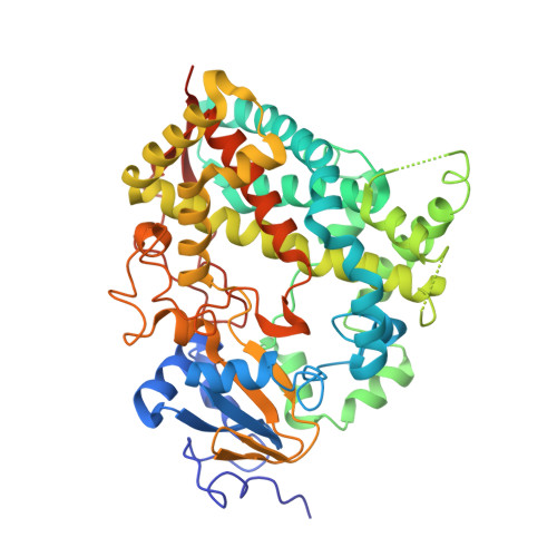
5A1P

Crystal structure of cytochrome P450 3A4 bound to progesterone and citrate


Experimental Data Snapshot

  • Method: X-RAY DIFFRACTION
  • Resolution: 2.50 Å
  • R-Value Free: 0.278 
  • R-Value Work: 0.191 
  • R-Value Observed: 0.195 

Starting Model: experimental
View more details

wwPDB Validation   3D Report Full Report


Ligand Structure Quality Assessment 


This is version 1.2 of the entry. See complete history


Literature

Anion-Dependent Stimulation of Cyp3A4 Monooxygenase.

Sevrioukova, I.F.Poulos, T.L.

(2015) Biochemistry 54: 4083

  • DOI: https://doi.org/10.1021/acs.biochem.5b00510
  • Primary Citation of Related Structures:  
    5A1P, 5A1R

  • PubMed Abstract: 

    We co-crystallized human cytochrome P450 3A4 (CYP3A4) with progesterone (PRG) under two different conditions, but the resulting complexes contained only one PRG molecule bound to the previously identified peripheral site. A novel feature in one of our structures is a citrate ion, originating from the crystallization solution. The citrate-binding site is located in an area where the N-terminus splits from the protein core and, thus, is suitable for the interaction with the anionic phospholipids of the microsomal membrane. We investigated how citrate affects the function of a soluble CYP3A4 monooxygenase system consisting of equimolar amounts of CYP3A4 and cytochrome P450 reductase (CPR). Citrate was found to affect the properties of both redox partners and stimulated their catalytic activities in a concentration-dependent manner via a complex mechanism. CYP3A4-substrate binding, reduction of CPR with NADPH, and interflavin and interprotein electron transfer were identified as citrate-sensitive steps. Comparative analysis of various negatively charged organic compounds indicated that, in addition to alterations caused by changes in ionic strength, anions modulate the properties of CYP3A4 and CPR through specific anion-protein interactions. Our results help to better understand previous observations and provide new mechanistic insights into CYP3A4 function.


  • Organizational Affiliation

    Departments of †Molecular Biology and Biochemistry, ‡Chemistry, and §Pharmaceutical Sciences, University of California, Irvine, California 92697-3900, United States.


Macromolecules
Find similar proteins by:  (by identity cutoff)  |  3D Structure
Entity ID: 1
MoleculeChains Sequence LengthOrganismDetailsImage
CYTOCHROME P450 3A4487Homo sapiensMutation(s): 0 
EC: 1.14.13.157 (PDB Primary Data), 1.14.13.32 (PDB Primary Data), 1.14.13.67 (PDB Primary Data), 1.14.13.9 (PDB Primary Data), 1.14.14.73 (UniProt), 1.14.14.1 (UniProt), 1.14.14.56 (UniProt), 1.14.14.55 (UniProt)
UniProt & NIH Common Fund Data Resources
Find proteins for P08684 (Homo sapiens)
Explore P08684 
Go to UniProtKB:  P08684
PHAROS:  P08684
GTEx:  ENSG00000160868 
Entity Groups  
Sequence Clusters30% Identity50% Identity70% Identity90% Identity95% Identity100% Identity
UniProt GroupP08684
Sequence Annotations
Expand
  • Reference Sequence
Experimental Data & Validation

Experimental Data

  • Method: X-RAY DIFFRACTION
  • Resolution: 2.50 Å
  • R-Value Free: 0.278 
  • R-Value Work: 0.191 
  • R-Value Observed: 0.195 
  • Space Group: I 2 2 2
Unit Cell:
Length ( Å )Angle ( ˚ )
a = 77.34α = 90
b = 102.17β = 90
c = 125.59γ = 90
Software Package:
Software NamePurpose
PHENIXrefinement
MOSFLMdata reduction
SCALAdata scaling
PHASERphasing

Structure Validation

View Full Validation Report


Ligand Structure Quality Assessment 


Entry History 

Deposition Data

Revision History  (Full details and data files)

  • Version 1.0: 2015-06-24
    Type: Initial release
  • Version 1.1: 2015-07-22
    Changes: Database references
  • Version 1.2: 2024-01-10
    Changes: Data collection, Database references, Derived calculations, Other, Refinement description