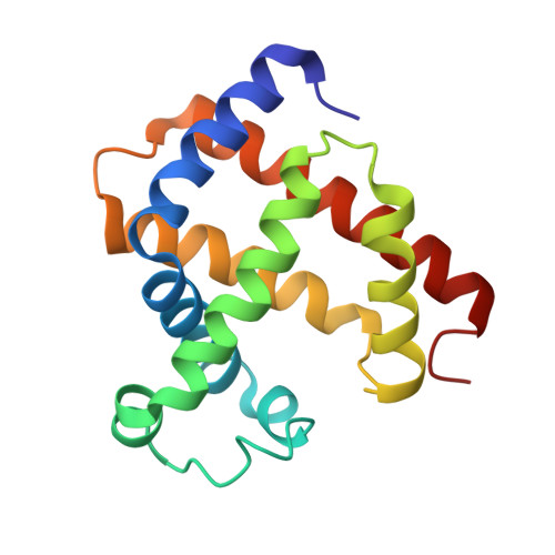
5AZQ

Crystal structure of cyano-cobalt(III) tetradehydrocorrin in the heme pocket of horse heart myoglobin


Experimental Data Snapshot

  • Method: X-RAY DIFFRACTION
  • Resolution: 1.40 Å
  • R-Value Free: 0.189 
  • R-Value Work: 0.128 
  • R-Value Observed: 0.131 

wwPDB Validation   3D Report Full Report


Ligand Structure Quality Assessment 


This is version 1.3 of the entry. See complete history


Literature

Crystal Structures and Coordination Behavior of Aqua- and Cyano-Co(III) Tetradehydrocorrins in the Heme Pocket of Myoglobin

Morita, Y.Oohora, K.Mizohata, E.Sawada, A.Kamachi, T.Yoshizawa, K.Inoue, T.Hayashi, T.

(2016) Inorg Chem 55: 1287-1295

  • DOI: https://doi.org/10.1021/acs.inorgchem.5b02598
  • Primary Citation of Related Structures:  
    5AZQ, 5AZR

  • PubMed Abstract: 

    Myoglobins reconstituted with aqua- and cyano-Co(III) tetradehydrocorrins, rMb(Co(III)(OH2)(TDHC)) and rMb(Co(III)(CN)(TDHC)), respectively, were prepared and investigated as models of a cobalamin-dependent enzyme. The former protein was obtained by oxidation of rMb(Co(II)(TDHC)) with K3[Fe(CN)6]. The cyanide-coordinated Co(III) species in the latter protein was prepared by ligand exchange of rMb(Co(III)(OH2)(TDHC)) with exogenous cyanide upon addition of KCN. The X-ray crystallographic study reveals the hexacoordinated structures of rMb(Co(III)(OH)(TDHC)) and rMb(Co(III)(CN)(TDHC)) at 1.20 and 1.40 Å resolution, respectively. The (13)C NMR chemical shifts of the cyanide in rMb(Co(III)(CN)(TDHC)) were determined to be 108.6 and 110.6 ppm. IR measurements show that the cyanide of rMb(Co(III)(CN)(TDHC)) has a stretching frequency peak at 2151 cm(-1) which is higher than that of cyanocobalamin. The (13)C NMR and IR measurements indicate weaker coordination of the cyanide to Co(III)(TDHC) relative to cobalamin, a vitamin B12 derivative. Thus, the extent of π-back-donation from the cobalt ion to the cyanide ion is lower in rMb(Co(III)(CN)(TDHC)). Furthermore, the pK(1/2) values of rMb(Co(III)(OH2)(TDHC)) and rMb(Co(III)(CN)(TDHC)) were determined by a pH titration experiment to be 3.2 and 5.5, respectively, indicating that the cyanide ligation weakens the Co-N(His93) bond. Theoretical calculations also demonstrate that the axial ligand exchange from water to cyanide elongates the Co-N(axial) bond with a decrease in the bond dissociation energy. Taken together, the cyano-Co(III) tetradehydrocorrin in myoglobin is appropriate for investigation as a structural analogue of methylcobalamin, a key intermediate in methionine synthase reaction.


  • Organizational Affiliation

    Department of Applied Chemistry, Graduate School of Engineering, Osaka University , Suita 565-0871, Japan.


Macromolecules
Find similar proteins by:  (by identity cutoff)  |  3D Structure
Entity ID: 1
MoleculeChains Sequence LengthOrganismDetailsImage
Myoglobin153Equus caballusMutation(s): 0 
EC: 1.11.1 (UniProt), 1.7 (UniProt)
UniProt
Find proteins for P68082 (Equus caballus)
Explore P68082 
Go to UniProtKB:  P68082
Entity Groups  
Sequence Clusters30% Identity50% Identity70% Identity90% Identity95% Identity100% Identity
UniProt GroupP68082
Sequence Annotations
Expand
  • Reference Sequence
Small Molecules
Ligands 4 Unique
IDChains Name / Formula / InChI Key2D Diagram3D Interactions
J1R
Query on J1R

Download Ideal Coordinates CCD File 
B [auth A](1R,19R) cobalt tetradehydrocorrin
C33 H37 Co N4 O4
DBXMOQHUTDEXAY-ZNBLAKOKSA-M
J1S
Query on J1S

Download Ideal Coordinates CCD File 
C [auth A](1S,19S) cobalt tetradehydrocorrin
C33 H37 Co N4 O4
DBXMOQHUTDEXAY-MLGYITDRSA-M
SO4
Query on SO4

Download Ideal Coordinates CCD File 
E [auth A],
F [auth A]
SULFATE ION
O4 S
QAOWNCQODCNURD-UHFFFAOYSA-L
CYN
Query on CYN

Download Ideal Coordinates CCD File 
D [auth A]CYANIDE ION
C N
XFXPMWWXUTWYJX-UHFFFAOYSA-N
Experimental Data & Validation

Experimental Data

  • Method: X-RAY DIFFRACTION
  • Resolution: 1.40 Å
  • R-Value Free: 0.189 
  • R-Value Work: 0.128 
  • R-Value Observed: 0.131 
  • Space Group: P 1 21 1
Unit Cell:
Length ( Å )Angle ( ˚ )
a = 34.774α = 90
b = 28.808β = 105.64
c = 63.258γ = 90
Software Package:
Software NamePurpose
REFMACrefinement
HKL-2000data reduction
Cootmodel building
PHASERphasing

Structure Validation

View Full Validation Report


Ligand Structure Quality Assessment 


Entry History & Funding Information

Deposition Data


Funding OrganizationLocationGrant Number
JSPS KAKENHIJapan15K18487

Revision History  (Full details and data files)

  • Version 1.0: 2016-01-20
    Type: Initial release
  • Version 1.1: 2016-02-17
    Changes: Database references
  • Version 1.2: 2020-02-26
    Changes: Data collection, Database references, Derived calculations
  • Version 1.3: 2024-03-20
    Changes: Data collection, Database references