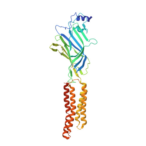
5CFB

Crystal Structure of Human Glycine Receptor alpha-3 Bound to Strychnine


Experimental Data Snapshot

  • Method: X-RAY DIFFRACTION
  • Resolution: 3.04 Å
  • R-Value Free: 0.283 
  • R-Value Work: 0.260 
  • R-Value Observed: 0.262 

Starting Model: experimental
View more details

wwPDB Validation   3D Report Full Report


Ligand Structure Quality Assessment 


This is version 2.2 of the entry. See complete history


Literature

Crystal structure of human glycine receptor-alpha 3 bound to antagonist strychnine.

Huang, X.Chen, H.Michelsen, K.Schneider, S.Shaffer, P.L.

(2015) Nature 526: 277-280

  • DOI: https://doi.org/10.1038/nature14972
  • Primary Citation of Related Structures:  
    5CFB

  • PubMed Abstract: 

    Neurotransmitter-gated ion channels of the Cys-loop receptor family are essential mediators of fast neurotransmission throughout the nervous system and are implicated in many neurological disorders. Available X-ray structures of prokaryotic and eukaryotic Cys-loop receptors provide tremendous insights into the binding of agonists, the subsequent opening of the ion channel, and the mechanism of channel activation. Yet the mechanism of inactivation by antagonists remains unknown. Here we present a 3.0 Å X-ray structure of the human glycine receptor-α3 homopentamer in complex with a high affinity, high-specificity antagonist, strychnine. Our structure allows us to explore in detail the molecular recognition of antagonists. Comparisons with previous structures reveal a mechanism for antagonist-induced inactivation of Cys-loop receptors, involving an expansion of the orthosteric binding site in the extracellular domain that is coupled to closure of the ion pore in the transmembrane domain.


  • Organizational Affiliation

    Department of Molecular Structure and Characterization, Amgen Inc., 360 Binney Street, Cambridge, Massachusetts 02142, USA.


Macromolecules
Find similar proteins by:  (by identity cutoff)  |  3D Structure
Entity ID: 1
MoleculeChains Sequence LengthOrganismDetailsImage
Glycine receptor subunit alpha-3,Glycine receptor subunit alpha-3
A, B, C, D, E
362Homo sapiensMutation(s): 0 
Gene Names: GLRA3
Membrane Entity: Yes 
UniProt & NIH Common Fund Data Resources
Find proteins for O75311 (Homo sapiens)
Explore O75311 
Go to UniProtKB:  O75311
PHAROS:  O75311
GTEx:  ENSG00000145451 
Entity Groups  
Sequence Clusters30% Identity50% Identity70% Identity90% Identity95% Identity100% Identity
UniProt GroupO75311
Glycosylation
Glycosylation Sites: 1Go to GlyGen: O75311-1
Sequence Annotations
Expand
  • Reference Sequence
Oligosaccharides

Help

Entity ID: 2
MoleculeChains Length2D Diagram Glycosylation3D Interactions
2-acetamido-2-deoxy-beta-D-glucopyranose-(1-4)-2-acetamido-2-deoxy-beta-D-glucopyranose
F, G
2N-Glycosylation
Glycosylation Resources
GlyTouCan:  G42666HT
GlyCosmos:  G42666HT
GlyGen:  G42666HT
Small Molecules
Ligands 2 Unique
IDChains Name / Formula / InChI Key2D Diagram3D Interactions
SY9
Query on SY9

Download Ideal Coordinates CCD File 
H [auth A],
K [auth B],
L [auth C],
M [auth D],
N [auth E]
STRYCHNINE
C21 H22 N2 O2
QMGVPVSNSZLJIA-FVWCLLPLSA-N
NAG
Query on NAG

Download Ideal Coordinates CCD File 
I [auth A],
J [auth B],
O [auth E]
2-acetamido-2-deoxy-beta-D-glucopyranose
C8 H15 N O6
OVRNDRQMDRJTHS-FMDGEEDCSA-N
Experimental Data & Validation

Experimental Data

  • Method: X-RAY DIFFRACTION
  • Resolution: 3.04 Å
  • R-Value Free: 0.283 
  • R-Value Work: 0.260 
  • R-Value Observed: 0.262 
  • Space Group: P 21 21 21
Unit Cell:
Length ( Å )Angle ( ˚ )
a = 140.242α = 90
b = 143.198β = 90
c = 180.054γ = 90
Software Package:
Software NamePurpose
REFMACrefinement
SCALEPACKdata scaling
PDB_EXTRACTdata extraction
PHASERphasing

Structure Validation

View Full Validation Report


Ligand Structure Quality Assessment 


Entry History 

Deposition Data

Revision History  (Full details and data files)

  • Version 1.0: 2015-09-30
    Type: Initial release
  • Version 1.1: 2015-10-14
    Changes: Database references
  • Version 1.2: 2015-10-21
    Changes: Database references
  • Version 1.3: 2016-07-20
    Changes: Data collection
  • Version 1.4: 2017-11-22
    Changes: Derived calculations, Refinement description
  • Version 2.0: 2020-07-29
    Type: Remediation
    Reason: Carbohydrate remediation
    Changes: Atomic model, Data collection, Derived calculations, Structure summary
  • Version 2.1: 2023-09-27
    Changes: Data collection, Database references, Refinement description, Structure summary
  • Version 2.2: 2024-11-06
    Changes: Structure summary