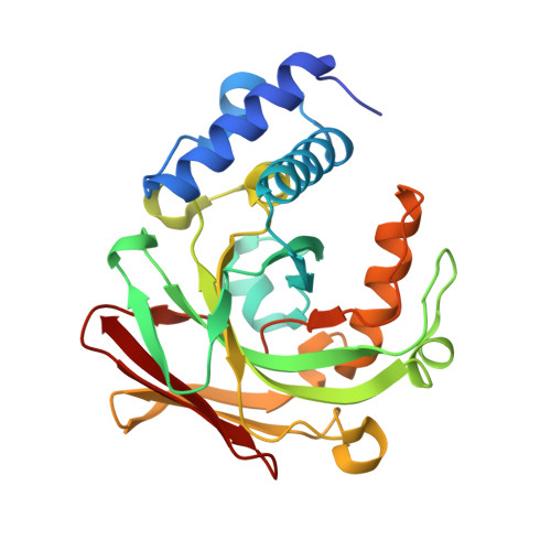
5D2K

4-oxalocrotonate decarboxylase from Pseudomonas putida G7 - complexed with magnesium and 2-oxoadipate


Experimental Data Snapshot

  • Method: X-RAY DIFFRACTION
  • Resolution: 1.57 Å
  • R-Value Free: 0.175 
  • R-Value Work: 0.150 
  • R-Value Observed: 0.152 

Starting Model: experimental
View more details

wwPDB Validation   3D Report Full Report


Ligand Structure Quality Assessment 


This is version 1.4 of the entry. See complete history


Literature

Crystal Structures of Apo and Liganded 4-Oxalocrotonate Decarboxylase Uncover a Structural Basis for the Metal-Assisted Decarboxylation of a Vinylogous beta-Keto Acid.

Guimaraes, S.L.Coitinho, J.B.Costa, D.M.Araujo, S.S.Whitman, C.P.Nagem, R.A.

(2016) Biochemistry 55: 2632-2645

  • DOI: https://doi.org/10.1021/acs.biochem.6b00050
  • Primary Citation of Related Structures:  
    5D2F, 5D2G, 5D2H, 5D2I, 5D2J, 5D2K

  • PubMed Abstract: 

    The enzymes in the catechol meta-fission pathway have been studied for more than 50 years in several species of bacteria capable of degrading a number of aromatic compounds. In a related pathway, naphthalene, a toxic polycyclic aromatic hydrocarbon, is fully degraded to intermediates of the tricarboxylic acid cycle by the soil bacteria Pseudomonas putida G7. In this organism, the 83 kb NAH7 plasmid carries several genes involved in this biotransformation process. One enzyme in this route, NahK, a 4-oxalocrotonate decarboxylase (4-OD), converts 2-oxo-3-hexenedioate to 2-hydroxy-2,4-pentadienoate using Mg(2+) as a cofactor. Efforts to study how 4-OD catalyzes this decarboxylation have been hampered because 4-OD is present in a complex with vinylpyruvate hydratase (VPH), which is the next enzyme in the same pathway. For the first time, a monomeric, stable, and active 4-OD has been expressed and purified in the absence of VPH. Crystal structures for NahK in the apo form and bonded with five substrate analogues were obtained using two distinct crystallization conditions. Analysis of the crystal structures implicates a lid domain in substrate binding and suggests roles for specific residues in a proposed reaction mechanism. In addition, we assign a possible function for the NahK N-terminal domain, which differs from most of the other members of the fumarylacetoacetate hydrolase superfamily. Although the structural basis for metal-dependent β-keto acid decarboxylases has been reported, this is the first structural report for that of a vinylogous β-keto acid decarboxylase and the first crystal structure of a 4-OD.


  • Organizational Affiliation

    Departamento de Bioquímica e Imunologia, Instituto de Ciências Biológicas, Universidade Federal de Minas Gerais , Belo Horizonte, 31270-901, Brazil.


Macromolecules
Find similar proteins by:  (by identity cutoff)  |  3D Structure
Entity ID: 1
MoleculeChains Sequence LengthOrganismDetailsImage
4-oxalocrotonate decarboxylase NahK
A, B
269Pseudomonas putidaMutation(s): 1 
Gene Names: nahK
EC: 4.1.1.77
UniProt
Find proteins for Q1XGK3 (Pseudomonas putida)
Explore Q1XGK3 
Go to UniProtKB:  Q1XGK3
Entity Groups  
Sequence Clusters30% Identity50% Identity70% Identity90% Identity95% Identity100% Identity
UniProt GroupQ1XGK3
Sequence Annotations
Expand
  • Reference Sequence
Small Molecules
Ligands 4 Unique
IDChains Name / Formula / InChI Key2D Diagram3D Interactions
OOG
Query on OOG

Download Ideal Coordinates CCD File 
D [auth A],
M [auth B]
2-OXOADIPIC ACID
C6 H8 O5
FGSBNBBHOZHUBO-UHFFFAOYSA-N
EDO
Query on EDO

Download Ideal Coordinates CCD File 
E [auth A]
F [auth A]
G [auth A]
H [auth A]
I [auth A]
E [auth A],
F [auth A],
G [auth A],
H [auth A],
I [auth A],
J [auth A],
K [auth A],
N [auth B],
O [auth B],
P [auth B],
Q [auth B],
R [auth B],
S [auth B]
1,2-ETHANEDIOL
C2 H6 O2
LYCAIKOWRPUZTN-UHFFFAOYSA-N
ACT
Query on ACT

Download Ideal Coordinates CCD File 
T [auth B]ACETATE ION
C2 H3 O2
QTBSBXVTEAMEQO-UHFFFAOYSA-M
MG
Query on MG

Download Ideal Coordinates CCD File 
C [auth A],
L [auth B]
MAGNESIUM ION
Mg
JLVVSXFLKOJNIY-UHFFFAOYSA-N
Experimental Data & Validation

Experimental Data

  • Method: X-RAY DIFFRACTION
  • Resolution: 1.57 Å
  • R-Value Free: 0.175 
  • R-Value Work: 0.150 
  • R-Value Observed: 0.152 
  • Space Group: P 1 21 1
Unit Cell:
Length ( Å )Angle ( ˚ )
a = 79.328α = 90
b = 44.411β = 118.68
c = 81.623γ = 90
Software Package:
Software NamePurpose
HKL-2000data reduction
HKL-2000data scaling
AMoREphasing
PHENIXrefinement

Structure Validation

View Full Validation Report


Ligand Structure Quality Assessment 


Entry History & Funding Information

Deposition Data


Funding OrganizationLocationGrant Number
CAPESBrazil--
FAPEMIGBrazilRDP-00174-10; EDT-0185-0.00-0; Rede-170/08
Brazilian National Council for Scientific and Technological Development (CNPq)Brazil482173/2010-6
VALE S.A.BrazilRDP-00174-10

Revision History  (Full details and data files)

  • Version 1.0: 2016-05-04
    Type: Initial release
  • Version 1.1: 2016-05-25
    Changes: Database references
  • Version 1.2: 2019-04-17
    Changes: Author supporting evidence, Data collection, Database references, Derived calculations
  • Version 1.3: 2020-01-08
    Changes: Author supporting evidence
  • Version 1.4: 2023-09-27
    Changes: Data collection, Database references, Derived calculations, Refinement description