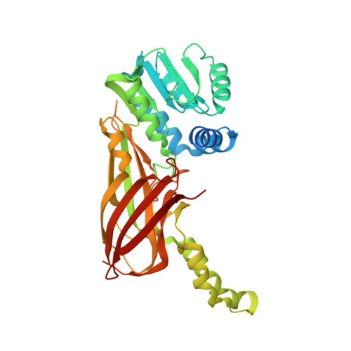
5EGS

Human PRMT6 with bound fragment-type inhibitor


Experimental Data Snapshot

  • Method: X-RAY DIFFRACTION
  • Resolution: 2.15 Å
  • R-Value Free: 0.246 
  • R-Value Work: 0.201 
  • R-Value Observed: 0.202 

Starting Model: experimental
View more details

wwPDB Validation   3D Report Full Report


Ligand Structure Quality Assessment 


This is version 1.2 of the entry. See complete history


Literature

Discovery of a Potent Class I Protein Arginine Methyltransferase Fragment Inhibitor.

Ferreira de Freitas, R.Eram, M.S.Szewczyk, M.M.Steuber, H.Smil, D.Wu, H.Li, F.Senisterra, G.Dong, A.Brown, P.J.Hitchcock, M.Moosmayer, D.Stegmann, C.M.Egner, U.Arrowsmith, C.Barsyte-Lovejoy, D.Vedadi, M.Schapira, M.

(2016) J Med Chem 59: 1176-1183

  • DOI: https://doi.org/10.1021/acs.jmedchem.5b01772
  • Primary Citation of Related Structures:  
    5EGS

  • PubMed Abstract: 

    Protein methyltransferases (PMTs) are a promising target class in oncology and other disease areas. They are composed of SET domain methyltransferases and structurally unrelated Rossman-fold enzymes that include protein arginine methyltransferases (PRMTs). In the absence of a well-defined medicinal chemistry tool-kit focused on PMTs, most current inhibitors were identified by screening large and diverse libraries of leadlike molecules. So far, no successful fragment-based approach was reported against this target class. Here, by deconstructing potent PRMT inhibitors, we find that chemical moieties occupying the substrate arginine-binding site can act as efficient fragment inhibitors. Screening a fragment library against PRMT6 produced numerous hits, including a 300 nM inhibitor (ligand efficiency of 0.56) that decreased global histone 3 arginine 2 methylation in cells, and can serve as a warhead for the development of PRMT chemical probes.


  • Organizational Affiliation

    Structural Genomics Consortium, University of Toronto , Toronto, ON M5G 1L7, Canada.


Macromolecules
Find similar proteins by:  (by identity cutoff)  |  3D Structure
Entity ID: 1
MoleculeChains Sequence LengthOrganismDetailsImage
Protein arginine N-methyltransferase 6
A, B, C, D
376Homo sapiensMutation(s): 0 
Gene Names: PRMT6HRMT1L6
EC: 2.1.1 (PDB Primary Data), 2.1.1.125 (PDB Primary Data), 2.1.1.319 (UniProt)
UniProt & NIH Common Fund Data Resources
Find proteins for Q96LA8 (Homo sapiens)
Explore Q96LA8 
Go to UniProtKB:  Q96LA8
PHAROS:  Q96LA8
GTEx:  ENSG00000198890 
Entity Groups  
Sequence Clusters30% Identity50% Identity70% Identity90% Identity95% Identity100% Identity
UniProt GroupQ96LA8
Sequence Annotations
Expand
  • Reference Sequence
Small Molecules
Ligands 2 Unique
IDChains Name / Formula / InChI Key2D Diagram3D Interactions
SAH
Query on SAH

Download Ideal Coordinates CCD File 
E [auth A],
H [auth B],
J [auth C],
M [auth D]
S-ADENOSYL-L-HOMOCYSTEINE
C14 H20 N6 O5 S
ZJUKTBDSGOFHSH-WFMPWKQPSA-N
5NR
Query on 5NR

Download Ideal Coordinates CCD File 
F [auth A]
G [auth A]
I [auth B]
K [auth C]
L [auth C]
F [auth A],
G [auth A],
I [auth B],
K [auth C],
L [auth C],
N [auth D]
2-[4-(phenylmethyl)piperidin-1-yl]ethanamine
C14 H22 N2
PCNDXYHWLSHXMV-UHFFFAOYSA-N
Experimental Data & Validation

Experimental Data

  • Method: X-RAY DIFFRACTION
  • Resolution: 2.15 Å
  • R-Value Free: 0.246 
  • R-Value Work: 0.201 
  • R-Value Observed: 0.202 
  • Space Group: P 1 21 1
Unit Cell:
Length ( Å )Angle ( ˚ )
a = 77.157α = 90
b = 135.616β = 98.91
c = 83.086γ = 90
Software Package:
Software NamePurpose
REFMACrefinement
XDSdata reduction
XSCALEdata scaling
PHASERphasing

Structure Validation

View Full Validation Report


Ligand Structure Quality Assessment 


Entry History 

Deposition Data

Revision History  (Full details and data files)

  • Version 1.0: 2016-02-03
    Type: Initial release
  • Version 1.1: 2016-06-22
    Changes: Database references
  • Version 1.2: 2024-01-10
    Changes: Data collection, Database references, Refinement description