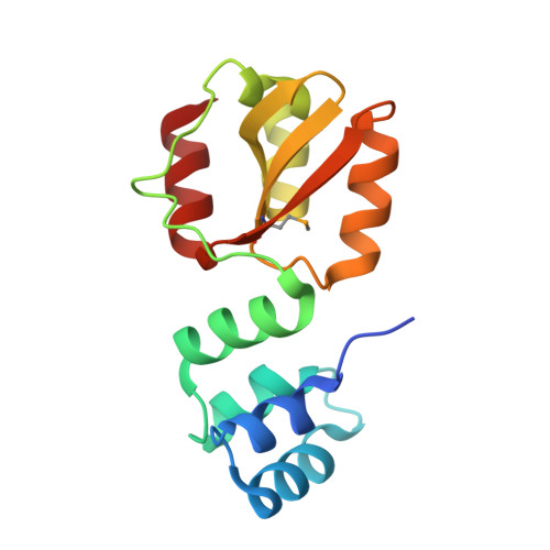
5HTL

Structure of MshE with cdg


Experimental Data Snapshot

  • Method: X-RAY DIFFRACTION
  • Resolution: 1.37 Å
  • R-Value Free: 0.221 
  • R-Value Work: 0.201 
  • R-Value Observed: 0.203 

wwPDB Validation   3D Report Full Report


Ligand Structure Quality Assessment 


This is version 1.2 of the entry. See complete history


Literature

Nucleotide binding by the widespread high-affinity cyclic di-GMP receptor MshEN domain.

Wang, Y.C.Chin, K.H.Tu, Z.L.He, J.Jones, C.J.Sanchez, D.Z.Yildiz, F.H.Galperin, M.Y.Chou, S.H.

(2016) Nat Commun 7: 12481-12481

  • DOI: https://doi.org/10.1038/ncomms12481
  • Primary Citation of Related Structures:  
    5HTL

  • PubMed Abstract: 

    C-di-GMP is a bacterial second messenger regulating various cellular functions. Many bacteria contain c-di-GMP-metabolizing enzymes but lack known c-di-GMP receptors. Recently, two MshE-type ATPases associated with bacterial type II secretion system and type IV pilus formation were shown to specifically bind c-di-GMP. Here we report crystal structure of the MshE N-terminal domain (MshEN1-145) from Vibrio cholerae in complex with c-di-GMP at a 1.37 Å resolution. This structure reveals a unique c-di-GMP-binding mode, featuring a tandem array of two highly conserved binding motifs, each comprising a 24-residue sequence RLGxx(L/V/I)(L/V/I)xxG(L/V/I)(L/V/I)xxxxLxxxLxxQ that binds half of the c-di-GMP molecule, primarily through hydrophobic interactions. Mutating these highly conserved residues markedly reduces c-di-GMP binding and biofilm formation by V. cholerae. This c-di-GMP-binding motif is present in diverse bacterial proteins exhibiting binding affinities ranging from 0.5 μM to as low as 14 nM. The MshEN domain contains the longest nucleotide-binding motif reported to date.


  • Organizational Affiliation

    Institute of Biochemistry, National Chung Hsing University, Taichung 40227, Taiwan, Republic of China.


Macromolecules
Find similar proteins by:  (by identity cutoff)  |  3D Structure
Entity ID: 1
MoleculeChains Sequence LengthOrganismDetailsImage
MSHA biogenesis protein MshE
A, B
145Vibrio choleraeMutation(s): 0 
Gene Names: epsE_3ERS013202_01172
UniProt
Find proteins for Q9KUV7 (Vibrio cholerae serotype O1 (strain ATCC 39315 / El Tor Inaba N16961))
Explore Q9KUV7 
Go to UniProtKB:  Q9KUV7
Entity Groups  
Sequence Clusters30% Identity50% Identity70% Identity90% Identity95% Identity100% Identity
UniProt GroupQ9KUV7
Sequence Annotations
Expand
  • Reference Sequence
Small Molecules
Ligands 1 Unique
IDChains Name / Formula / InChI Key2D Diagram3D Interactions
C2E
Query on C2E

Download Ideal Coordinates CCD File 
C [auth A],
D [auth B]
9,9'-[(2R,3R,3aS,5S,7aR,9R,10R,10aS,12S,14aR)-3,5,10,12-tetrahydroxy-5,12-dioxidooctahydro-2H,7H-difuro[3,2-d:3',2'-j][1,3,7,9,2,8]tetraoxadiphosphacyclododecine-2,9-diyl]bis(2-amino-1,9-dihydro-6H-purin-6-one)
C20 H24 N10 O14 P2
PKFDLKSEZWEFGL-MHARETSRSA-N
Modified Residues  1 Unique
IDChains TypeFormula2D DiagramParent
MSE
Query on MSE
A, B
L-PEPTIDE LINKINGC5 H11 N O2 SeMET
Experimental Data & Validation

Experimental Data

  • Method: X-RAY DIFFRACTION
  • Resolution: 1.37 Å
  • R-Value Free: 0.221 
  • R-Value Work: 0.201 
  • R-Value Observed: 0.203 
  • Space Group: P 21 21 21
Unit Cell:
Length ( Å )Angle ( ˚ )
a = 52.852α = 90
b = 61.862β = 90
c = 75.621γ = 90
Software Package:
Software NamePurpose
PHENIXrefinement
HKL-2000data reduction
HKL-2000data scaling
PHASERphasing

Structure Validation

View Full Validation Report


Ligand Structure Quality Assessment 


Entry History 

Deposition Data

Revision History  (Full details and data files)

  • Version 1.0: 2016-10-05
    Type: Initial release
  • Version 1.1: 2016-10-12
    Changes: Database references
  • Version 1.2: 2024-10-09
    Changes: Data collection, Database references, Derived calculations, Structure summary