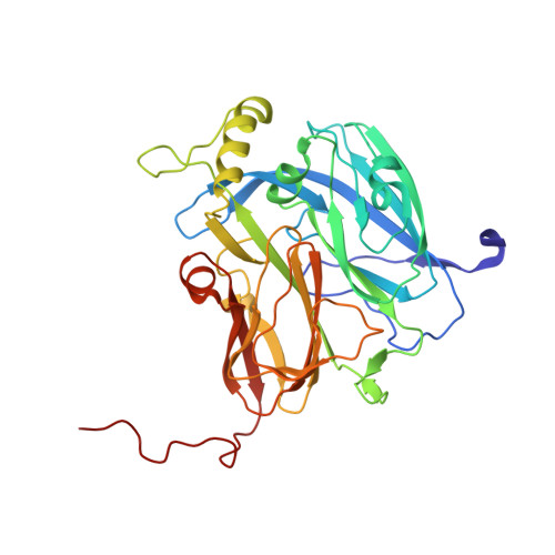
5I6M

Crystal Structure of Copper Nitrite Reductase at 100K after 7.59 MGy


Experimental Data Snapshot

  • Method: X-RAY DIFFRACTION
  • Resolution: 1.09 Å
  • R-Value Free: 0.146 
  • R-Value Work: 0.127 
  • R-Value Observed: 0.128 

Starting Model: experimental
View more details

wwPDB Validation   3D Report Full Report


This is version 1.5 of the entry. See complete history


Literature

Serial crystallography captures enzyme catalysis in copper nitrite reductase at atomic resolution from one crystal.

Horrell, S.Antonyuk, S.V.Eady, R.R.Hasnain, S.S.Hough, M.A.Strange, R.W.

(2016) IUCrJ 3: 271-281

  • DOI: https://doi.org/10.1107/S205225251600823X
  • Primary Citation of Related Structures:  
    5I6K, 5I6L, 5I6M, 5I6N, 5I6O, 5I6P

  • PubMed Abstract: 

    Relating individual protein crystal structures to an enzyme mechanism remains a major and challenging goal for structural biology. Serial crystallography using multiple crystals has recently been reported in both synchrotron-radiation and X-ray free-electron laser experiments. In this work, serial crystallography was used to obtain multiple structures serially from one crystal (MSOX) to study in crystallo enzyme catalysis. Rapid, shutterless X-ray detector technology on a synchrotron MX beamline was exploited to perform low-dose serial crystallography on a single copper nitrite reductase crystal, which survived long enough for 45 consecutive 100 K X-ray structures to be collected at 1.07-1.62 Å resolution, all sampled from the same crystal volume. This serial crystallography approach revealed the gradual conversion of the substrate bound at the catalytic type 2 Cu centre from nitrite to nitric oxide, following reduction of the type 1 Cu electron-transfer centre by X-ray-generated solvated electrons. Significant, well defined structural rearrangements in the active site are evident in the series as the enzyme moves through its catalytic cycle, namely nitrite reduction, which is a vital step in the global denitrification process. It is proposed that such a serial crystallography approach is widely applicable for studying any redox or electron-driven enzyme reactions from a single protein crystal. It can provide a 'catalytic reaction movie' highlighting the structural changes that occur during enzyme catalysis. The anticipated developments in the automation of data analysis and modelling are likely to allow seamless and near-real-time analysis of such data on-site at some of the powerful synchrotron crystallographic beamlines.


  • Organizational Affiliation

    School of Biological Sciences, University of Essex , Wivenhoe Park, Colchester CO4 3SQ, England.


Macromolecules
Find similar proteins by:  (by identity cutoff)  |  3D Structure
Entity ID: 1
MoleculeChains Sequence LengthOrganismDetailsImage
Copper-containing nitrite reductase332Achromobacter cycloclastesMutation(s): 0 
Gene Names: nirK
EC: 1.7.2.1
UniProt
Find proteins for P25006 (Achromobacter cycloclastes)
Explore P25006 
Go to UniProtKB:  P25006
Entity Groups  
Sequence Clusters30% Identity50% Identity70% Identity90% Identity95% Identity100% Identity
UniProt GroupP25006
Sequence Annotations
Expand
  • Reference Sequence
Small Molecules
Ligands 5 Unique
IDChains Name / Formula / InChI Key2D Diagram3D Interactions
MLI
Query on MLI

Download Ideal Coordinates CCD File 
E [auth A]MALONATE ION
C3 H2 O4
OFOBLEOULBTSOW-UHFFFAOYSA-L
CU
Query on CU

Download Ideal Coordinates CCD File 
B [auth A],
C [auth A]
COPPER (II) ION
Cu
JPVYNHNXODAKFH-UHFFFAOYSA-N
ACT
Query on ACT

Download Ideal Coordinates CCD File 
D [auth A]ACETATE ION
C2 H3 O2
QTBSBXVTEAMEQO-UHFFFAOYSA-M
NO2
Query on NO2

Download Ideal Coordinates CCD File 
F [auth A]
H [auth A]
I [auth A]
J [auth A]
K [auth A]
F [auth A],
H [auth A],
I [auth A],
J [auth A],
K [auth A],
L [auth A],
M [auth A]
NITRITE ION
N O2
IOVCWXUNBOPUCH-UHFFFAOYSA-M
NO
Query on NO

Download Ideal Coordinates CCD File 
G [auth A]NITRIC OXIDE
N O
ODUCDPQEXGNKDN-UHFFFAOYSA-N
Experimental Data & Validation

Experimental Data

  • Method: X-RAY DIFFRACTION
  • Resolution: 1.09 Å
  • R-Value Free: 0.146 
  • R-Value Work: 0.127 
  • R-Value Observed: 0.128 
  • Space Group: P 21 3
Unit Cell:
Length ( Å )Angle ( ˚ )
a = 95.155α = 90
b = 95.155β = 90
c = 95.155γ = 90
Software Package:
Software NamePurpose
REFMACrefinement
Aimlessdata scaling
PDB_EXTRACTdata extraction
XDSdata reduction
REFMACphasing

Structure Validation

View Full Validation Report


Entry History & Funding Information

Deposition Data


Funding OrganizationLocationGrant Number
Biotechnology and Biological Sciences Research CouncilUnited KingdomBB/M020924/1
Biotechnology and Biological Sciences Research CouncilUnited KingdomBB/M022714/1

Revision History  (Full details and data files)

  • Version 1.0: 2016-07-13
    Type: Initial release
  • Version 1.1: 2016-07-27
    Changes: Database references
  • Version 1.2: 2016-09-21
    Changes: Database references
  • Version 1.3: 2016-09-28
    Changes: Database references
  • Version 1.4: 2017-08-30
    Changes: Author supporting evidence, Data collection
  • Version 1.5: 2024-01-10
    Changes: Data collection, Database references, Derived calculations, Refinement description