5ITE

2.2-Angstrom in meso crystal structure of Haloquadratum Walsbyi Bacteriorhodopsin (HwBR) from Octylglucoside (OG) Detergent Micelles


Experimental Data Snapshot

  • Method: X-RAY DIFFRACTION
  • Resolution: 2.18 Å
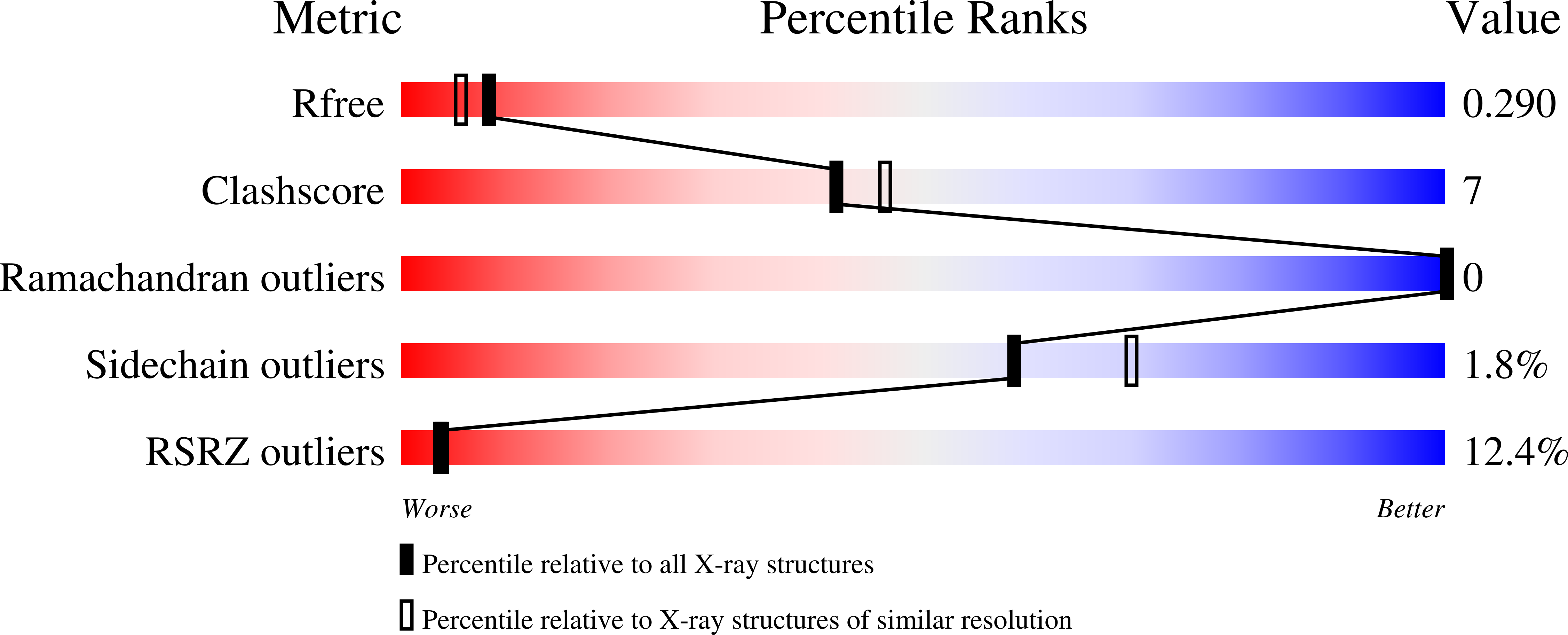
  • R-Value Free: 0.289 
  • R-Value Work: 0.259 
  • R-Value Observed: 0.261 

Starting Model: experimental
View more details

wwPDB Validation   3D Report Full Report


Ligand Structure Quality Assessment 


This is version 1.4 of the entry. See complete history


Literature

Crystallogenesis of Membrane Proteins Mediated by Polymer-Bounded Lipid Nanodiscs.

Broecker, J.Eger, B.T.Ernst, O.P.

(2017) Structure 25: 384-392

  • DOI: https://doi.org/10.1016/j.str.2016.12.004
  • Primary Citation of Related Structures:  
    5ITC, 5ITE

  • PubMed Abstract: 

    For some membrane proteins, detergent-mediated solubilization compromises protein stability and functionality, often impairing biophysical and structural analyses. Hence, membrane-protein structure determination is a continuing bottleneck in the field of protein crystallography. Here, as an alternative to approaches mediated by conventional detergents, we report the crystallogenesis of a recombinantly produced membrane protein that never left a lipid bilayer environment. We used styrene-maleic acid (SMA) copolymers to solubilize lipid-embedded proteins into SMA nanodiscs, purified these discs by affinity and size-exclusion chromatography, and transferred proteins into the lipidic cubic phase (LCP) for in meso crystallization. The 2.0-Å structure of an α-helical seven-transmembrane microbial rhodopsin thus obtained is of high quality and virtually identical to the 2.2-Å structure obtained from traditional detergent-based purification and subsequent LCP crystallization.


  • Organizational Affiliation

    Structural Neurobiology, Department of Biochemistry, University of Toronto, 1 King's College Circle, Toronto, ON M5S 1A8, Canada. Electronic address: [email protected].


Macromolecules
Find similar proteins by:  (by identity cutoff)  |  3D Structure
Entity ID: 1
MoleculeChains Sequence LengthOrganismDetailsImage
Bacteriorhodopsin-I
A, B, C
268Haloquadratum walsbyi DSM 16790Mutation(s): 0 
Gene Names: bop1bopIHQ_1014A
Membrane Entity: Yes 
UniProt
Find proteins for Q18DH8 (Haloquadratum walsbyi (strain DSM 16790 / HBSQ001))
Explore Q18DH8 
Go to UniProtKB:  Q18DH8
Entity Groups  
Sequence Clusters30% Identity50% Identity70% Identity90% Identity95% Identity100% Identity
UniProt GroupQ18DH8
Sequence Annotations
Expand
  • Reference Sequence
Small Molecules
Ligands 3 Unique
IDChains Name / Formula / InChI Key2D Diagram3D Interactions
OLC
Query on OLC

Download Ideal Coordinates CCD File 
F [auth A]
H [auth B]
I [auth B]
J [auth B]
M [auth C]
F [auth A],
H [auth B],
I [auth B],
J [auth B],
M [auth C],
N [auth C]
(2R)-2,3-dihydroxypropyl (9Z)-octadec-9-enoate
C21 H40 O4
RZRNAYUHWVFMIP-GDCKJWNLSA-N
OLB
Query on OLB

Download Ideal Coordinates CCD File 
E [auth A],
K [auth B],
O [auth C]
(2S)-2,3-dihydroxypropyl (9Z)-octadec-9-enoate
C21 H40 O4
RZRNAYUHWVFMIP-QJRAZLAKSA-N
RET
Query on RET

Download Ideal Coordinates CCD File 
D [auth A],
G [auth B],
L [auth C]
RETINAL
C20 H28 O
NCYCYZXNIZJOKI-OVSJKPMPSA-N
Experimental Data & Validation

Experimental Data

  • Method: X-RAY DIFFRACTION
  • Resolution: 2.18 Å
  • R-Value Free: 0.289 
  • R-Value Work: 0.259 
  • R-Value Observed: 0.261 
  • Space Group: C 1 2 1
Unit Cell:
Length ( Å )Angle ( ˚ )
a = 106.442α = 90
b = 61.565β = 116.8
c = 119.689γ = 90
Software Package:
Software NamePurpose
PHENIXrefinement
autoPROCdata scaling
PHASERphasing

Structure Validation

View Full Validation Report


Ligand Structure Quality Assessment 


Entry History & Funding Information

Deposition Data


Funding OrganizationLocationGrant Number
Government of CanadaCanadaCanada Excellence Research Chair Program
German Research Foundation (DFG)GermanyBR 5124/1-1

Revision History  (Full details and data files)

  • Version 1.0: 2017-01-25
    Type: Initial release
  • Version 1.1: 2017-02-22
    Changes: Database references
  • Version 1.2: 2017-11-22
    Changes: Refinement description
  • Version 1.3: 2023-09-27
    Changes: Data collection, Database references, Refinement description
  • Version 1.4: 2024-11-06
    Changes: Structure summary