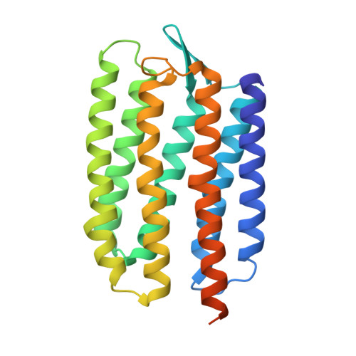
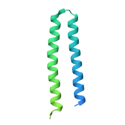
5JJE

Structure of the SRII/HtrII Complex in I212121 space group ("U" shape)


Experimental Data Snapshot

  • Method: X-RAY DIFFRACTION
  • Resolution: 1.90 Å
  • R-Value Free: 0.218 
  • R-Value Work: 0.197 
  • R-Value Observed: 0.198 

Starting Model: experimental
View more details

wwPDB Validation   3D Report Full Report


Ligand Structure Quality Assessment 


This is version 1.4 of the entry. See complete history


Literature

New Insights on Signal Propagation by Sensory Rhodopsin II/Transducer Complex.

Ishchenko, A.Round, E.Borshchevskiy, V.Grudinin, S.Gushchin, I.Klare, J.P.Remeeva, A.Polovinkin, V.Utrobin, P.Balandin, T.Engelhard, M.Buldt, G.Gordeliy, V.

(2017) Sci Rep 7: 41811-41811

  • DOI: https://doi.org/10.1038/srep41811
  • Primary Citation of Related Structures:  
    5JJE, 5JJF, 5JJJ, 5JJN

  • PubMed Abstract: 

    The complex of two membrane proteins, sensory rhodopsin II (NpSRII) with its cognate transducer (NpHtrII), mediates negative phototaxis in halobacteria N. pharaonis. Upon light activation NpSRII triggers a signal transduction chain homologous to the two-component system in eubacterial chemotaxis. Here we report on crystal structures of the ground and active M-state of the complex in the space group I2 1 2 1 2 1 . We demonstrate that the relative orientation of symmetrical parts of the dimer is parallel ("U"-shaped) contrary to the gusset-like ("V"-shaped) form of the previously reported structures of the NpSRII/NpHtrII complex in the space group P2 1 2 1 2, although the structures of the monomers taken individually are nearly the same. Computer modeling of the HAMP domain in the obtained "V"- and "U"-shaped structures revealed that only the "U"-shaped conformation allows for tight interactions of the receptor with the HAMP domain. This is in line with existing data and supports biological relevance of the "U" shape in the ground state. We suggest that the "V"-shaped structure may correspond to the active state of the complex and transition from the "U" to the "V"-shape of the receptor-transducer complex can be involved in signal transduction from the receptor to the signaling domain of NpHtrII.


  • Organizational Affiliation

    Institute of Complex Systems (ICS), ICS-6: Structural Biochemistry, Research Centre Jülich, 52425 Jülich, Germany.


Macromolecules
Find similar proteins by:  (by identity cutoff)  |  3D Structure
Entity ID: 1
MoleculeChains Sequence LengthOrganismDetailsImage
Sensory rhodopsin-2248Natronomonas pharaonisMutation(s): 0 
Gene Names: sop2sopII
Membrane Entity: Yes 
UniProt
Find proteins for P42196 (Natronomonas pharaonis)
Explore P42196 
Go to UniProtKB:  P42196
Entity Groups  
Sequence Clusters30% Identity50% Identity70% Identity90% Identity95% Identity100% Identity
UniProt GroupP42196
Sequence Annotations
Expand
  • Reference Sequence
Find similar proteins by:  (by identity cutoff)  |  3D Structure
Entity ID: 2
MoleculeChains Sequence LengthOrganismDetailsImage
Sensory rhodopsin II transducer163Natronomonas pharaonisMutation(s): 0 
Gene Names: htr2htrII
Membrane Entity: Yes 
UniProt
Find proteins for P42259 (Natronomonas pharaonis)
Explore P42259 
Go to UniProtKB:  P42259
Entity Groups  
Sequence Clusters30% Identity50% Identity70% Identity90% Identity95% Identity100% Identity
UniProt GroupP42259
Sequence Annotations
Expand
  • Reference Sequence
Small Molecules
Ligands 3 Unique
IDChains Name / Formula / InChI Key2D Diagram3D Interactions
BOG
Query on BOG

Download Ideal Coordinates CCD File 
D [auth A]octyl beta-D-glucopyranoside
C14 H28 O6
HEGSGKPQLMEBJL-RKQHYHRCSA-N
RET
Query on RET

Download Ideal Coordinates CCD File 
C [auth A]RETINAL
C20 H28 O
NCYCYZXNIZJOKI-OVSJKPMPSA-N
LFA
Query on LFA

Download Ideal Coordinates CCD File 
E [auth A]
F [auth A]
G [auth A]
H [auth A]
I [auth A]
E [auth A],
F [auth A],
G [auth A],
H [auth A],
I [auth A],
J [auth A],
K [auth A],
L [auth A],
M [auth A],
N [auth B],
O [auth B],
P [auth B]
EICOSANE
C20 H42
CBFCDTFDPHXCNY-UHFFFAOYSA-N
Experimental Data & Validation

Experimental Data

  • Method: X-RAY DIFFRACTION
  • Resolution: 1.90 Å
  • R-Value Free: 0.218 
  • R-Value Work: 0.197 
  • R-Value Observed: 0.198 
  • Space Group: I 21 21 21
Unit Cell:
Length ( Å )Angle ( ˚ )
a = 49.642α = 90
b = 113.661β = 90
c = 125.995γ = 90
Software Package:
Software NamePurpose
PHENIXrefinement
MOSFLMdata reduction
SCALAdata scaling
MOLREPphasing

Structure Validation

View Full Validation Report


Ligand Structure Quality Assessment 


Entry History & Funding Information

Deposition Data


Funding OrganizationLocationGrant Number
Russian Science FoundationRussian Federation14-14-00995
French National Research AgencyFranceANR-10-INSB-05-02

Revision History  (Full details and data files)

  • Version 1.0: 2017-02-15
    Type: Initial release
  • Version 1.1: 2017-09-06
    Changes: Advisory, Author supporting evidence
  • Version 1.2: 2020-07-29
    Type: Remediation
    Reason: Carbohydrate remediation
    Changes: Data collection, Derived calculations, Structure summary
  • Version 1.3: 2024-01-10
    Changes: Advisory, Data collection, Database references, Refinement description, Structure summary
  • Version 1.4: 2024-11-06
    Changes: Structure summary