5LFG

X-ray structure of a new fully ligated carbomonoxy form of Trematomus newnesi hemoglobin (Hb1TnCO).


Experimental Data Snapshot

  • Method: X-RAY DIFFRACTION
  • Resolution: 1.94 Å
  • R-Value Free: 0.245 
  • R-Value Work: 0.181 

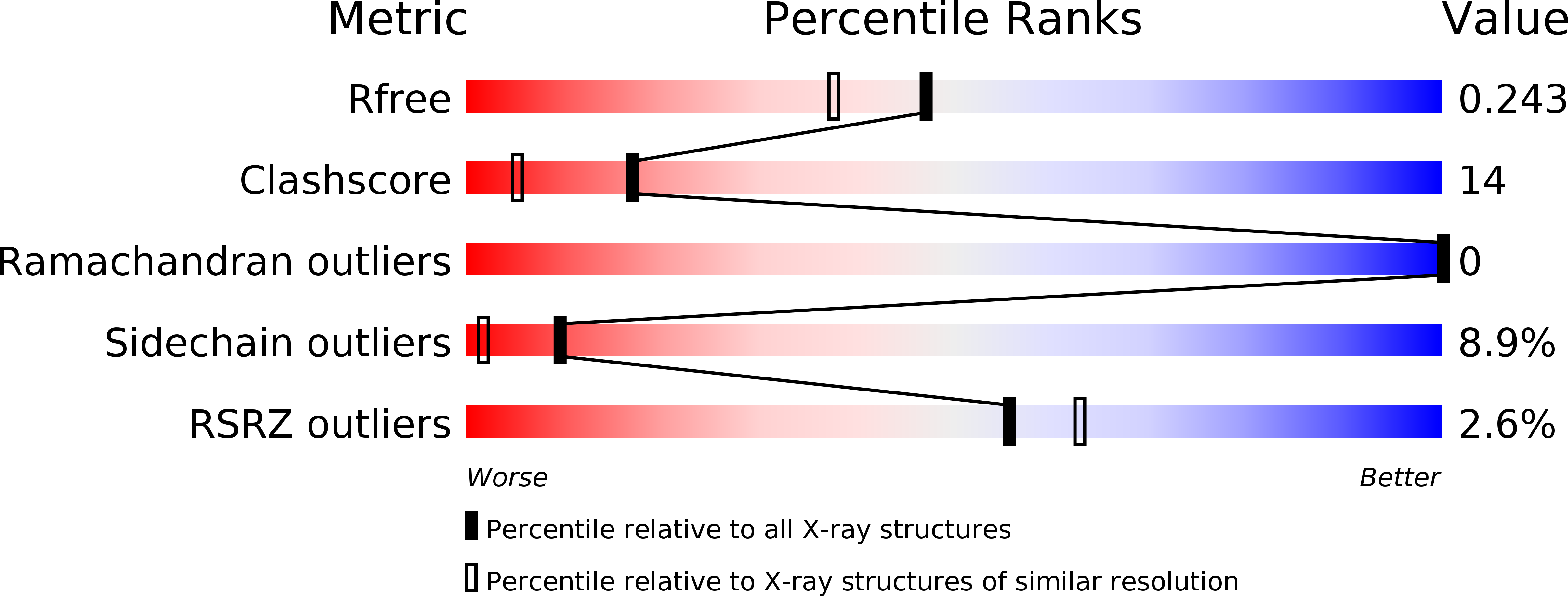
wwPDB Validation   3D Report Full Report


Ligand Structure Quality Assessment 


This is version 1.1 of the entry. See complete history


Literature

Fine Sampling of the RT Quaternary-Structure Transition of a Tetrameric Hemoglobin.

Vitagliano, L.Mazzarella, L.Merlino, A.Vergara, A.

(2017) Chemistry 23: 605-613

  • DOI: https://doi.org/10.1002/chem.201603421
  • Primary Citation of Related Structures:  
    5LFG

  • PubMed Abstract: 

    Although the end points of the functional transitions of tetrameric hemoglobins (Hbs) have been well characterized, atomic-resolution data on R-T intermediate states are extremely limited. Herein, the X-ray structures of two independent tetramers of the fully ligated carbomonoxy form of Trematomus newnesi hemoglobin (Hb1Tn) within the same crystal are described. These structures show peculiar features in the heme pocket, EF corner, and tertiary/quaternary structure. Distal histidine side chains have a propensity to swing out of the heme pocket and thus allow compression of the EF corner. In this rotameric state, the distal His group does not interact with the CO ligand, consistent with FTIR spectra recorded in solution. At the quaternary-structure level, one tetramer is an intermediate R-T state, whereas the other assumes a T-like structure. Altogether, the structures of these tetramers provide the best available atomic-level picture of the R→T transition of vertebrate Hbs.


  • Organizational Affiliation

    Institute of Biostructures and Biomaging, CNR, Via Mezzocannone 16, 80134, Napoli, Italy.


Macromolecules
Find similar proteins by:  (by identity cutoff)  |  3D Structure
Entity ID: 1
MoleculeChains Sequence LengthOrganismDetailsImage
Hemoglobin subunit alpha-1
A, C
143Trematomus newnesiMutation(s): 1 
UniProt
Find proteins for P45718 (Trematomus newnesi)
Explore P45718 
Go to UniProtKB:  P45718
Entity Groups  
Sequence Clusters30% Identity50% Identity70% Identity90% Identity95% Identity100% Identity
UniProt GroupP45718
Sequence Annotations
Expand
  • Reference Sequence
Find similar proteins by:  (by identity cutoff)  |  3D Structure
Entity ID: 2
MoleculeChains Sequence LengthOrganismDetailsImage
Hemoglobin subunit beta-1/2
B, D
146Trematomus newnesiMutation(s): 0 
UniProt
Find proteins for P45720 (Trematomus newnesi)
Explore P45720 
Go to UniProtKB:  P45720
Entity Groups  
Sequence Clusters30% Identity50% Identity70% Identity90% Identity95% Identity100% Identity
UniProt GroupP45720
Sequence Annotations
Expand
  • Reference Sequence
Small Molecules
Ligands 2 Unique
IDChains Name / Formula / InChI Key2D Diagram3D Interactions
HEM
Query on HEM

Download Ideal Coordinates CCD File 
F [auth A],
H [auth B],
J [auth C],
K [auth D]
PROTOPORPHYRIN IX CONTAINING FE
C34 H32 Fe N4 O4
KABFMIBPWCXCRK-RGGAHWMASA-L
CMO
Query on CMO

Download Ideal Coordinates CCD File 
E [auth A],
G [auth B],
I [auth C],
L [auth D]
CARBON MONOXIDE
C O
UGFAIRIUMAVXCW-UHFFFAOYSA-N
Experimental Data & Validation

Experimental Data

  • Method: X-RAY DIFFRACTION
  • Resolution: 1.94 Å
  • R-Value Free: 0.245 
  • R-Value Work: 0.181 
  • Space Group: C 1 2 1
Unit Cell:
Length ( Å )Angle ( ˚ )
a = 86.206α = 90
b = 87.256β = 101.81
c = 109.648γ = 90
Software Package:
Software NamePurpose
SHELXLrefinement
DENZOdata reduction
SCALEPACKdata scaling
AMoREphasing

Structure Validation

View Full Validation Report


Ligand Structure Quality Assessment 


Entry History 

Deposition Data

Revision History  (Full details and data files)

  • Version 1.0: 2017-08-09
    Type: Initial release
  • Version 1.1: 2024-10-16
    Changes: Data collection, Database references, Derived calculations, Structure summary