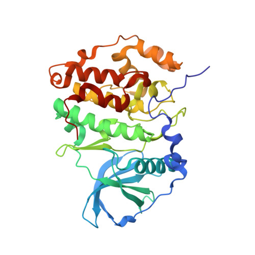
5N9N

Crystal structure of human Protein kinase CK2 catalytic subunit in complex with the ATP-competitive, tight-binding dibenzofuran inhibitor TF85 (4a)


Experimental Data Snapshot

  • Method: X-RAY DIFFRACTION
  • Resolution: 1.84 Å
  • R-Value Free: 0.201 
  • R-Value Work: 0.167 
  • R-Value Observed: 0.169 

Starting Model: experimental
View more details

wwPDB Validation   3D Report Full Report


Ligand Structure Quality Assessment 


This is version 2.1 of the entry. See complete history


Literature

A pi-Halogen Bond of Dibenzofuranones with the Gatekeeper Phe113 in Human Protein Kinase CK2 Leads to Potent Tight Binding Inhibitors.

Schnitzler, A.Gratz, A.Bollacke, A.Weyrich, M.Kucklander, U.Wunsch, B.Gotz, C.Niefind, K.Jose, J.

(2018) Pharmaceuticals (Basel) 11

  • DOI: https://doi.org/10.3390/ph11010023
  • Primary Citation of Related Structures:  
    5N9K, 5N9L, 5N9N

  • PubMed Abstract: 

    Human protein kinase CK2 is an emerging target for neoplastic diseases. Potent lead structures for human CK2 inhibitors are derived from dibenzofuranones. Two new derivatives, 7,9-dichloro-1,2-dihydro-8-hydroxy-4-[(4-methoxyphenylamino)-methylene]dibenzo[ b , d ]furan-3(2 H )-one ( 4a ) and ( E )-1,3-dichloro-6-[(4-methoxyphenylimino)-methyl]dibenzo[ b , d ]furan-2,7-diol ( 5 ) were tested for inhibition of CK2 and induction of apoptosis in LNCaP cells. Both turned out to be tight binding inhibitors, with IC 50 values of 7 nM ( 4a ) and 5 nM ( 5 ) and an apparent K i value of 0.4 nM for both. Compounds 4a and 5 reduced cellular CK2 activity, indicating cell permeability. Cell viability was substantially impaired in LNCaP cells, as well as apoptosis was induced, which was not appearing in non-neoplastic ARPE-19 cells. Co-crystallization of 4a and 5 revealed an unexpected π -halogen bond of the chloro substituent at C9 with the gatekeeper amino acid Phe113, leading to an inverted binding mode in comparison to parent compound 4b , with the Cl at C6 instead, which was co-crystallized as a control. This indicates that the position of the chloro substituent on ring A of the dibenzofuran scaffold is responsible for an inversion of the binding mode that enhances potency.


  • Organizational Affiliation

    Institut für Biochemie, Department für Chemie, Universität zu Köln, Zülpicher Straße 47, D-50674 Köln, Germany. [email protected].


Macromolecules
Find similar proteins by:  (by identity cutoff)  |  3D Structure
Entity ID: 1
MoleculeChains Sequence LengthOrganismDetailsImage
Casein kinase II subunit alpha335Homo sapiensMutation(s): 0 
Gene Names: CSNK2A1CK2A1
EC: 2.7.11.1
UniProt & NIH Common Fund Data Resources
Find proteins for P68400 (Homo sapiens)
Explore P68400 
Go to UniProtKB:  P68400
PHAROS:  P68400
GTEx:  ENSG00000101266 
Entity Groups  
Sequence Clusters30% Identity50% Identity70% Identity90% Identity95% Identity100% Identity
UniProt GroupP68400
Sequence Annotations
Expand
  • Reference Sequence
Small Molecules
Ligands 3 Unique
IDChains Name / Formula / InChI Key2D Diagram3D Interactions
KC5
Query on KC5

Download Ideal Coordinates CCD File 
B [auth A](4~{Z})-7,9-bis(chloranyl)-4-[[(4-methoxyphenyl)amino]methylidene]-8-oxidanyl-1,2-dihydrodibenzofuran-3-one
C20 H15 Cl2 N O4
GOSOFAONCKLXHN-UKTHLTGXSA-N
GOL
Query on GOL

Download Ideal Coordinates CCD File 
H [auth A],
I [auth A],
J [auth A],
K [auth A],
L [auth A]
GLYCEROL
C3 H8 O3
PEDCQBHIVMGVHV-UHFFFAOYSA-N
ACT
Query on ACT

Download Ideal Coordinates CCD File 
C [auth A],
D [auth A],
E [auth A],
F [auth A],
G [auth A]
ACETATE ION
C2 H3 O2
QTBSBXVTEAMEQO-UHFFFAOYSA-M
Experimental Data & Validation

Experimental Data

  • Method: X-RAY DIFFRACTION
  • Resolution: 1.84 Å
  • R-Value Free: 0.201 
  • R-Value Work: 0.167 
  • R-Value Observed: 0.169 
  • Space Group: P 1 21 1
Unit Cell:
Length ( Å )Angle ( ˚ )
a = 57.477α = 90
b = 45.505β = 110.97
c = 63.489γ = 90
Software Package:
Software NamePurpose
PHENIXrefinement
XDSdata reduction
Aimlessdata scaling
PHASERphasing

Structure Validation

View Full Validation Report


Ligand Structure Quality Assessment 


Entry History 

Deposition Data

Revision History  (Full details and data files)

  • Version 1.0: 2018-02-28
    Type: Initial release
  • Version 1.1: 2018-03-07
    Changes: Database references
  • Version 1.2: 2018-04-18
    Changes: Data collection, Database references
  • Version 1.3: 2018-04-25
    Changes: Data collection, Database references
  • Version 2.0: 2019-05-22
    Changes: Atomic model, Data collection, Derived calculations, Non-polymer description, Structure summary
  • Version 2.1: 2024-01-17
    Changes: Data collection, Database references, Refinement description