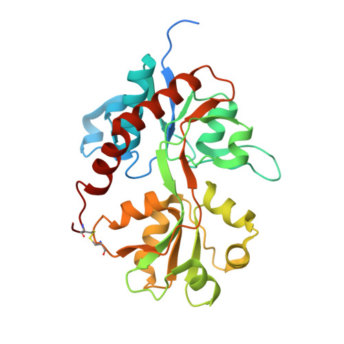
5NS9

Crystal structure of the GluA2 LBD (L483Y-N754S-L758V) in complex with glutamate


Experimental Data Snapshot

  • Method: X-RAY DIFFRACTION
  • Resolution: 1.44 Å
  • R-Value Free: 0.205 
  • R-Value Work: 0.147 
  • R-Value Observed: 0.150 

Starting Model: experimental
View more details

wwPDB Validation   3D Report Full Report


This is version 1.4 of the entry. See complete history


Literature

Unitary Properties of AMPA Receptors with Reduced Desensitization.

Zhang, W.Eibl, C.Weeks, A.M.Riva, I.Li, Y.J.Plested, A.J.R.Howe, J.R.

(2017) Biophys J 113: 2218-2235

  • DOI: https://doi.org/10.1016/j.bpj.2017.07.030
  • Primary Citation of Related Structures:  
    5NS9

  • PubMed Abstract: 

    Wild-type AMPA receptors display a characteristic rapidly desensitizing phenotype. Many studies point to the dimer interface between pairs of extracellular ligand binding domains as the key region controlling the rate at which the receptors desensitize. However, mutations at the extracellular end of the pore-forming regions (near the putative ion channel gate) have also been shown to alter desensitization. Here we report the behavior of single GluA4 receptors carrying one of two mutations that greatly reduce desensitization at the level of ensemble currents: the dimer interface mutation L484Y and the Lurcher mutation (A623T, GluA4-Lc) in the extracellular end of M3 (the second true transmembrane helix). Analysis of unitary currents in patches with just one active receptor showed that each mutation greatly prolongs bursts of openings without prolonging the apparent duration of individual openings. Each mutation decreases the frequency with which individual receptors visit desensitized states, but both mutant receptors still desensitize multiple times per second. Cyclothiazide (CTZ) reduced desensitization of wild-type receptors and both types of mutant receptor. Analysis of shut-time distributions revealed a form of short-lived desensitization that was resistant to CTZ and was especially prominent for GluA4-Lc receptors. Despite reducing desensitization of GluA4 L484Y receptors, CTZ decreased the amplitude of ensemble currents through GluA2 and GluA4 LY receptor mutants. Single-channel analysis and comparison of the GluA2 L483Y ligand binding domain dimer in complex with glutamate with and without CTZ is consistent with the conclusion that CTZ binding to the dimer interface prevents effects of the LY mutation to modulate receptor activation, resulting in a reduction in the prevalence of large-conductance substates that accounts for the decrease in ensemble current amplitudes. Together, the results show that similar nondesensitizing AMPA-receptor phenotypes of population currents can arise from distinct underlying molecular mechanisms that produce different types of unitary activity.


  • Organizational Affiliation

    Department of Pharmacology, Institution of Chinese Integrative Medicine, Hebei Medical University, Shijiazhuang, Hebei, China; Department of Pharmacology, Yale University School of Medicine, New Haven, Connecticut. Electronic address: [email protected].


Macromolecules
Find similar proteins by:  (by identity cutoff)  |  3D Structure
Entity ID: 1
MoleculeChains Sequence LengthOrganismDetailsImage
Glutamate receptor 2,Glutamate receptor 2
A, B
292Rattus norvegicusMutation(s): 3 
Gene Names: Gria2Glur2
UniProt
Find proteins for P19491 (Rattus norvegicus)
Explore P19491 
Go to UniProtKB:  P19491
Entity Groups  
Sequence Clusters30% Identity50% Identity70% Identity90% Identity95% Identity100% Identity
UniProt GroupP19491
Sequence Annotations
Expand
  • Reference Sequence
Small Molecules
Ligands 5 Unique
IDChains Name / Formula / InChI Key2D Diagram3D Interactions
1PE
Query on 1PE

Download Ideal Coordinates CCD File 
C [auth A],
D [auth A]
PENTAETHYLENE GLYCOL
C10 H22 O6
JLFNLZLINWHATN-UHFFFAOYSA-N
GLU
Query on GLU

Download Ideal Coordinates CCD File 
M [auth A],
R [auth B]
GLUTAMIC ACID
C5 H9 N O4
WHUUTDBJXJRKMK-VKHMYHEASA-N
PG0
Query on PG0

Download Ideal Coordinates CCD File 
E [auth A]2-(2-METHOXYETHOXY)ETHANOL
C5 H12 O3
SBASXUCJHJRPEV-UHFFFAOYSA-N
SO4
Query on SO4

Download Ideal Coordinates CCD File 
F [auth A]
G [auth A]
H [auth A]
I [auth A]
N [auth B]
F [auth A],
G [auth A],
H [auth A],
I [auth A],
N [auth B],
O [auth B]
SULFATE ION
O4 S
QAOWNCQODCNURD-UHFFFAOYSA-L
NA
Query on NA

Download Ideal Coordinates CCD File 
J [auth A],
K [auth A],
L [auth A],
P [auth B],
Q [auth B]
SODIUM ION
Na
FKNQFGJONOIPTF-UHFFFAOYSA-N
Experimental Data & Validation

Experimental Data

  • Method: X-RAY DIFFRACTION
  • Resolution: 1.44 Å
  • R-Value Free: 0.205 
  • R-Value Work: 0.147 
  • R-Value Observed: 0.150 
  • Space Group: P 21 21 2
Unit Cell:
Length ( Å )Angle ( ˚ )
a = 95.99α = 90
b = 121.297β = 90
c = 47.083γ = 90
Software Package:
Software NamePurpose
REFMACrefinement
XDSdata reduction
XSCALEdata scaling
PHASERphasing

Structure Validation

View Full Validation Report


Entry History & Funding Information

Deposition Data


Funding OrganizationLocationGrant Number
Austrian Science FundAustriaJ3682_B21
European Research Council647895

Revision History  (Full details and data files)

  • Version 1.0: 2017-09-13
    Type: Initial release
  • Version 1.1: 2017-11-29
    Changes: Database references
  • Version 1.2: 2019-10-16
    Changes: Data collection
  • Version 1.3: 2024-01-17
    Changes: Data collection, Database references, Derived calculations, Refinement description
  • Version 1.4: 2024-11-06
    Changes: Structure summary