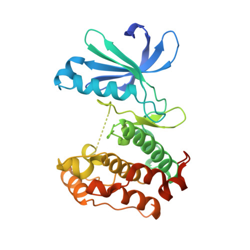
5ONE

Crystal structure of Aurora-A in complex with FMF-03-145-1 (compound 2)


Experimental Data Snapshot

  • Method: X-RAY DIFFRACTION
  • Resolution: 2.60 Å
  • R-Value Free: 0.260 
  • R-Value Work: 0.208 
  • R-Value Observed: 0.211 

Starting Model: experimental
View more details

wwPDB Validation   3D Report Full Report


Ligand Structure Quality Assessment 


This is version 1.2 of the entry. See complete history


Literature

Characterization of a highly selective inhibitor of the Aurora kinases.

Ferguson, F.M.Doctor, Z.M.Chaikuad, A.Sim, T.Kim, N.D.Knapp, S.Gray, N.S.

(2017) Bioorg Med Chem Lett 27: 4405-4408

  • DOI: https://doi.org/10.1016/j.bmcl.2017.08.016
  • Primary Citation of Related Structures:  
    5ONE

  • PubMed Abstract: 

    Aurora kinases play an essential role in mitosis and cell cycle regulation. In recent years Aurora kinases have proved popular cancer targets and many inhibitors have been developed. The majority of these clinical candidates are multi-targeted, rendering them inappropriate as tools for studying Aurora kinase mediated signaling. Here we report discovery of a highly selective inhibitor of Aurora kinases A, B and C, with potent cellular activity and minimal off-target activity (PLK4). The X-ray co-crystal structure of Aurora A in complex with compound 2 is reported, and provides insights into the structural determinants of ligand binding and selectivity.


  • Organizational Affiliation

    Department of Cancer Biology, Dana-Farber Cancer Institute, Department of Biological Chemistry and Molecular Pharmacology, Harvard Medical School, Boston, MA, USA.


Macromolecules
Find similar proteins by:  (by identity cutoff)  |  3D Structure
Entity ID: 1
MoleculeChains Sequence LengthOrganismDetailsImage
Aurora kinase A285Homo sapiensMutation(s): 0 
Gene Names: AURKAAIKAIRK1ARK1AURAAYK1BTAKIAK1STK15STK6
EC: 2.7.11.1
UniProt & NIH Common Fund Data Resources
Find proteins for O14965 (Homo sapiens)
Explore O14965 
Go to UniProtKB:  O14965
PHAROS:  O14965
GTEx:  ENSG00000087586 
Entity Groups  
Sequence Clusters30% Identity50% Identity70% Identity90% Identity95% Identity100% Identity
UniProt GroupO14965
Sequence Annotations
Expand
  • Reference Sequence
Small Molecules
Ligands 1 Unique
IDChains Name / Formula / InChI Key2D Diagram3D Interactions
9YQ
Query on 9YQ

Download Ideal Coordinates CCD File 
B [auth A]4-(propanoylamino)-~{N}-[4-[(5,8,11-trimethyl-6-oxidanylidene-pyrimido[4,5-b][1,4]benzodiazepin-2-yl)amino]phenyl]benzamide
C30 H29 N7 O3
KGXBCWANASZZQG-UHFFFAOYSA-N
Experimental Data & Validation

Experimental Data

  • Method: X-RAY DIFFRACTION
  • Resolution: 2.60 Å
  • R-Value Free: 0.260 
  • R-Value Work: 0.208 
  • R-Value Observed: 0.211 
  • Space Group: P 32 2 1
Unit Cell:
Length ( Å )Angle ( ˚ )
a = 86.765α = 90
b = 86.765β = 90
c = 77.103γ = 120
Software Package:
Software NamePurpose
REFMACrefinement
XDSdata reduction
Aimlessdata scaling
PHASERphasing

Structure Validation

View Full Validation Report


Ligand Structure Quality Assessment 


Entry History 

Deposition Data

Revision History  (Full details and data files)

  • Version 1.0: 2017-09-06
    Type: Initial release
  • Version 1.1: 2017-09-20
    Changes: Database references
  • Version 1.2: 2024-01-17
    Changes: Data collection, Database references, Refinement description