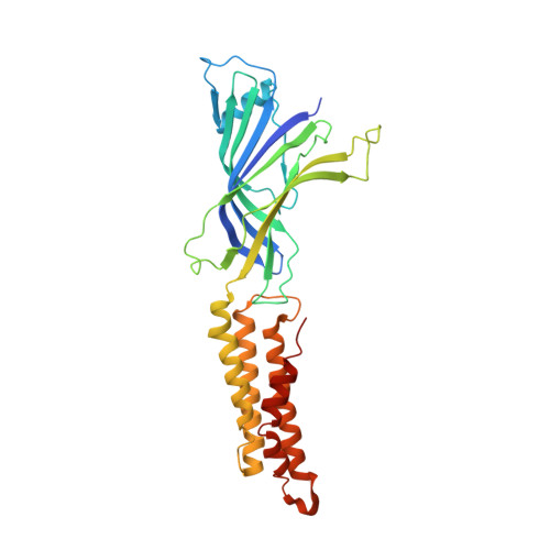
5SXU

X-ray structure of 2-bromoethanol bound to a pentameric ligand gated ion channel (ELIC) in a desensitized state


Experimental Data Snapshot

  • Method: X-RAY DIFFRACTION
  • Resolution: 3.10 Å
  • R-Value Free: 0.239 
  • R-Value Work: 0.200 
  • R-Value Observed: 0.202 

Starting Model: experimental
View more details

wwPDB Validation   3D Report Full Report


This is version 1.4 of the entry. See complete history


Literature

Structural Basis of Alcohol Inhibition of the Pentameric Ligand-Gated Ion Channel ELIC.

Chen, Q.Wells, M.M.Tillman, T.S.Kinde, M.N.Cohen, A.Xu, Y.Tang, P.

(2017) Structure 25: 180-187

  • DOI: https://doi.org/10.1016/j.str.2016.11.007
  • Primary Citation of Related Structures:  
    5SXU, 5SXV

  • PubMed Abstract: 

    The structural basis for alcohol modulation of neuronal pentameric ligand-gated ion channels (pLGICs) remains elusive. We determined an inhibitory mechanism of alcohol on the pLGIC Erwinia chrysanthemi (ELIC) through direct binding to the pore. X-ray structures of ELIC co-crystallized with 2-bromoethanol, in both the absence and presence of agonist, reveal 2-bromoethanol binding in the pore near T237(6') and the extracellular domain (ECD) of each subunit at three different locations. Binding to the ECD does not appear to contribute to the inhibitory action of 2-bromoethanol and ethanol as indicated by the same functional responses of wild-type ELIC and mutants. In contrast, the ELIC-α1β3GABA A R chimera, replacing the ELIC transmembrane domain (TMD) with the TMD of α1β3GABA A R, is potentiated by 2-bromoethanol and ethanol. The results suggest a dominant role of the TMD in modulating alcohol effects. The X-ray structures and functional measurements support a pore-blocking mechanism for inhibitory action of short-chain alcohols.


  • Organizational Affiliation

    Department of Anesthesiology, University of Pittsburgh School of Medicine, Pittsburgh, PA 15260, USA.


Macromolecules
Find similar proteins by:  (by identity cutoff)  |  3D Structure
Entity ID: 1
MoleculeChains Sequence LengthOrganismDetailsImage
Gamma-aminobutyric-acid receptor subunit beta-1
A, B, C, D, E
A, B, C, D, E, F, G, H, I, J
322Dickeya dadantii 3937Mutation(s): 0 
Gene Names: Dda3937_00520
Membrane Entity: Yes 
UniProt
Find proteins for E0SJQ4 (Dickeya dadantii (strain 3937))
Explore E0SJQ4 
Go to UniProtKB:  E0SJQ4
Entity Groups  
Sequence Clusters30% Identity50% Identity70% Identity90% Identity95% Identity100% Identity
UniProt GroupE0SJQ4
Sequence Annotations
Expand
  • Reference Sequence
Small Molecules
Ligands 4 Unique
IDChains Name / Formula / InChI Key2D Diagram3D Interactions
MES
Query on MES

Download Ideal Coordinates CCD File 
AB [auth H]
FA [auth D]
FB [auth I]
KA [auth E]
KB [auth J]
AB [auth H],
FA [auth D],
FB [auth I],
KA [auth E],
KB [auth J],
P [auth A],
QA [auth F],
U [auth B],
VA [auth G],
Z [auth C]
2-(N-MORPHOLINO)-ETHANESULFONIC ACID
C6 H13 N O4 S
SXGZJKUKBWWHRA-UHFFFAOYSA-N
BRJ
Query on BRJ

Download Ideal Coordinates CCD File 
BA [auth D]
BB [auth I]
CA [auth D]
CB [auth I]
DA [auth D]
BA [auth D],
BB [auth I],
CA [auth D],
CB [auth I],
DA [auth D],
DB [auth I],
HA [auth E],
HB [auth J],
IA [auth E],
IB [auth J],
JA [auth E],
JB [auth J],
K [auth A],
L [auth A],
LA [auth F],
M [auth A],
MA [auth F],
N [auth A],
NA [auth F],
OA [auth F],
Q [auth B],
R [auth B],
RA [auth G],
S [auth B],
SA [auth G],
TA [auth G],
V [auth C],
W [auth C],
WA [auth H],
X [auth C],
XA [auth H],
YA [auth H]
2-BROMOETHANOL
C2 H5 Br O
LDLCZOVUSADOIV-UHFFFAOYSA-N
3CN
Query on 3CN

Download Ideal Coordinates CCD File 
EA [auth D]
EB [auth I]
GA [auth E]
GB [auth J]
O [auth A]
EA [auth D],
EB [auth I],
GA [auth E],
GB [auth J],
O [auth A],
PA [auth F],
T [auth B],
UA [auth G],
Y [auth C],
ZA [auth H]
3-AMINOPROPANE
C3 H9 N
WGYKZJWCGVVSQN-UHFFFAOYSA-N
CL
Query on CL

Download Ideal Coordinates CCD File 
AA [auth C],
LB [auth J]
CHLORIDE ION
Cl
VEXZGXHMUGYJMC-UHFFFAOYSA-M
Experimental Data & Validation

Experimental Data

  • Method: X-RAY DIFFRACTION
  • Resolution: 3.10 Å
  • R-Value Free: 0.239 
  • R-Value Work: 0.200 
  • R-Value Observed: 0.202 
  • Space Group: P 1 21 1
Unit Cell:
Length ( Å )Angle ( ˚ )
a = 105.903α = 90
b = 266.791β = 107.13
c = 111.109γ = 90
Software Package:
Software NamePurpose
PHENIXrefinement
XDSdata scaling
Aimlessdata scaling
PHENIXphasing

Structure Validation

View Full Validation Report


Entry History & Funding Information

Deposition Data


Funding OrganizationLocationGrant Number
National Institutes of Health/National Institute of General Medical Sciences (NIH/NIGMS)United StatesR01GM066358
National Institutes of Health/National Institute of General Medical Sciences (NIH/NIGMS)United StatesR01GM056257
National Institutes of Health/National Institute of General Medical Sciences (NIH/NIGMS)United StatesT32GM075770
National Institutes of Health/National Institute of General Medical Sciences (NIH/NIGMS)United StatesR37GM049202

Revision History  (Full details and data files)

  • Version 1.0: 2017-06-21
    Type: Initial release
  • Version 1.1: 2017-09-13
    Changes: Author supporting evidence
  • Version 1.2: 2017-11-22
    Changes: Refinement description
  • Version 1.3: 2019-12-25
    Changes: Author supporting evidence
  • Version 1.4: 2023-10-04
    Changes: Data collection, Database references, Refinement description