5UJC

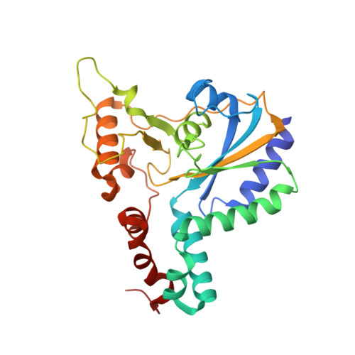
Crystal structure of a C.elegans B12-trafficking protein CblC, a human MMACHC homologue


Experimental Data Snapshot

  • Method: X-RAY DIFFRACTION
  • Resolution: 1.35 Å
  • R-Value Free: 0.163 
  • R-Value Work: 0.132 
  • R-Value Observed: 0.134 

Starting Model: experimental
View more details

wwPDB Validation   3D Report Full Report


Ligand Structure Quality Assessment 


This is version 1.4 of the entry. See complete history


Literature

Coordination chemistry controls the thiol oxidase activity of the B12-trafficking protein CblC.

Li, Z.Shanmuganathan, A.Ruetz, M.Yamada, K.Lesniak, N.A.Krautler, B.Brunold, T.C.Koutmos, M.Banerjee, R.

(2017) J Biol Chem 292: 9733-9744

  • DOI: https://doi.org/10.1074/jbc.M117.788554
  • Primary Citation of Related Structures:  
    5UJC

  • PubMed Abstract: 

    The cobalamin or B 12 cofactor supports sulfur and one-carbon metabolism and the catabolism of odd-chain fatty acids, branched-chain amino acids, and cholesterol. CblC is a B 12 -processing enzyme involved in an early cytoplasmic step in the cofactor-trafficking pathway. It catalyzes the glutathione (GSH)-dependent dealkylation of alkylcobalamins and the reductive decyanation of cyanocobalamin. CblC from Caenorhabditis elegans ( ce CblC) also exhibits a robust thiol oxidase activity, converting reduced GSH to oxidized GSSG with concomitant scrubbing of ambient dissolved O 2 The mechanism of thiol oxidation catalyzed by ce CblC is not known. In this study, we demonstrate that novel coordination chemistry accessible to ce CblC-bound cobalamin supports its thiol oxidase activity via a glutathionyl-cobalamin intermediate. Deglutathionylation of glutathionyl-cobalamin by a second molecule of GSH yields GSSG. The crystal structure of ce CblC provides insights into how architectural differences at the α- and β-faces of cobalamin promote the thiol oxidase activity of ce CblC but mute it in wild-type human CblC. The R161G and R161Q mutations in human CblC unmask its latent thiol oxidase activity and are correlated with increased cellular oxidative stress disease. In summary, we have uncovered key architectural features in the cobalamin-binding pocket that support unusual cob(II)alamin coordination chemistry and enable the thiol oxidase activity of ce CblC.


  • Organizational Affiliation

    From the Department of Biological Chemistry, University of Michigan Medical Center, Ann Arbor, Michigan 48109-0600.


Macromolecules
Find similar proteins by:  (by identity cutoff)  |  3D Structure
Entity ID: 1
MoleculeChains Sequence LengthOrganismDetailsImage
MMACHC-like protein270Caenorhabditis elegansMutation(s): 0 
Gene Names: cblc-1ZK546.17
EC: 1.16.1
UniProt
Find proteins for Q7Z144 (Caenorhabditis elegans)
Explore Q7Z144 
Go to UniProtKB:  Q7Z144
Entity Groups  
Sequence Clusters30% Identity50% Identity70% Identity90% Identity95% Identity100% Identity
UniProt GroupQ7Z144
Sequence Annotations
Expand
  • Reference Sequence
Small Molecules
Ligands 4 Unique
IDChains Name / Formula / InChI Key2D Diagram3D Interactions
COB
Query on COB

Download Ideal Coordinates CCD File 
B [auth A]CO-METHYLCOBALAMIN
C63 H91 Co N13 O14 P
BMOGZGJBQIUQJG-BYLSPCLQSA-N
TAR
Query on TAR

Download Ideal Coordinates CCD File 
E [auth A]D(-)-TARTARIC ACID
C4 H6 O6
FEWJPZIEWOKRBE-LWMBPPNESA-N
TLA
Query on TLA

Download Ideal Coordinates CCD File 
C [auth A],
D [auth A]
L(+)-TARTARIC ACID
C4 H6 O6
FEWJPZIEWOKRBE-JCYAYHJZSA-N
GOL
Query on GOL

Download Ideal Coordinates CCD File 
F [auth A]GLYCEROL
C3 H8 O3
PEDCQBHIVMGVHV-UHFFFAOYSA-N
Experimental Data & Validation

Experimental Data

  • Method: X-RAY DIFFRACTION
  • Resolution: 1.35 Å
  • R-Value Free: 0.163 
  • R-Value Work: 0.132 
  • R-Value Observed: 0.134 
  • Space Group: P 21 21 21
Unit Cell:
Length ( Å )Angle ( ˚ )
a = 53.008α = 90
b = 66.089β = 90
c = 92.826γ = 90
Software Package:
Software NamePurpose
REFMACrefinement
XDSdata reduction
Aimlessdata scaling
PHASERphasing

Structure Validation

View Full Validation Report


Ligand Structure Quality Assessment 


Entry History & Funding Information

Deposition Data


Funding OrganizationLocationGrant Number
American Heart AssociationUnited States13SDG14560056

Revision History  (Full details and data files)

  • Version 1.0: 2017-05-03
    Type: Initial release
  • Version 1.1: 2017-05-10
    Changes: Database references
  • Version 1.2: 2017-06-28
    Changes: Database references
  • Version 1.3: 2017-11-01
    Changes: Author supporting evidence
  • Version 1.4: 2023-10-04
    Changes: Data collection, Database references, Refinement description