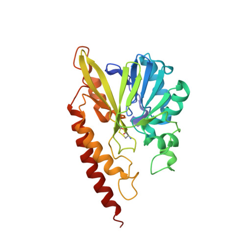
5WCM

Crystal structure of the complex between class B3 beta-lactamase BJP-1 and 4-nitrobenzene-sulfonamide - new refinement


Experimental Data Snapshot

  • Method: X-RAY DIFFRACTION
  • Resolution: 1.20 Å
  • R-Value Free: 0.160 
  • R-Value Work: 0.132 
  • R-Value Observed: 0.133 

Starting Model: experimental
View more details

wwPDB Validation   3D Report Full Report


Ligand Structure Quality Assessment 


This is version 1.4 of the entry. See complete history


Literature

High-resolution crystal structure of the subclass B3 metallo-beta-lactamase BJP-1: rational basis for substrate specificity and interaction with sulfonamides.

Docquier, J.D.Benvenuti, M.Calderone, V.Stoczko, M.Menciassi, N.Rossolini, G.M.Mangani, S.

(2010) Antimicrob Agents Chemother 54: 4343-4351

  • DOI: https://doi.org/10.1128/AAC.00409-10
  • Primary Citation of Related Structures:  
    3LVZ, 5WCM

  • PubMed Abstract: 

    Metallo-β-lactamases (MBLs) are important enzymatic factors in resistance to β-lactam antibiotics that show important structural and functional heterogeneity. BJP-1 is a subclass B3 MBL determinant produced by Bradyrhizobium japonicum that exhibits interesting properties. BJP-1, like CAU-1 of Caulobacter vibrioides, overall poorly recognizes β-lactam substrates and shows an unusual substrate profile compared to other MBLs. In order to understand the structural basis of these properties, the crystal structure of BJP-1 was obtained at 1.4-Å resolution. This revealed significant differences in the conformation and locations of the active-site loops, determining a rather narrow active site and the presence of a unique N-terminal helix bearing Phe-31, whose side chain binds in the active site and represents an obstacle for β-lactam substrate binding. In order to probe the potential of sulfonamides (known to inhibit various zinc-dependent enzymes) to bind in the active sites of MBLs, the structure of BJP-1 in complex with 4-nitrobenzenesulfonamide was also obtained (at 1.33-A resolution), thereby revealing the mode of interaction of these molecules in MBLs. Interestingly, sulfonamide binding resulted in the displacement of the side chain of Phe-31 from its hydrophobic binding pocket, where the benzene ring of the molecule is now found. These data further highlight the structural diversity shown by MBLs but also provide interesting insights in the structure-function relationships of these enzymes. More importantly, we provided the first structural observation of MBL interaction with sulfonamides, which might represent an interesting scaffold for the design of MBL inhibitors.


  • Organizational Affiliation

    Dipartimento di Biologia Molecolare, Laboratorio di Fisiologia e Biotecnologia dei Microrganismi, Università di Siena, I-53100 Siena, Italy.


Macromolecules
Find similar proteins by:  (by identity cutoff)  |  3D Structure
Entity ID: 1
MoleculeChains Sequence LengthOrganismDetailsImage
Blr6230 protein
A, B
263Bradyrhizobium diazoefficiens USDA 110Mutation(s): 0 
Gene Names: blr6230
UniProt
Find proteins for Q89GW5 (Bradyrhizobium diazoefficiens (strain JCM 10833 / BCRC 13528 / IAM 13628 / NBRC 14792 / USDA 110))
Explore Q89GW5 
Go to UniProtKB:  Q89GW5
Entity Groups  
Sequence Clusters30% Identity50% Identity70% Identity90% Identity95% Identity100% Identity
UniProt GroupQ89GW5
Sequence Annotations
Expand
  • Reference Sequence
Small Molecules
Ligands 2 Unique
IDChains Name / Formula / InChI Key2D Diagram3D Interactions
4NZ
Query on 4NZ

Download Ideal Coordinates CCD File 
I [auth A],
O [auth B]
4-nitrobenzenesulfonamide
C6 H6 N2 O4 S
QWKKYJLAUWFPDB-UHFFFAOYSA-N
ZN
Query on ZN

Download Ideal Coordinates CCD File 
C [auth A]
D [auth A]
E [auth A]
F [auth A]
G [auth A]
C [auth A],
D [auth A],
E [auth A],
F [auth A],
G [auth A],
H [auth A],
J [auth B],
K [auth B],
L [auth B],
M [auth B],
N [auth B]
ZINC ION
Zn
PTFCDOFLOPIGGS-UHFFFAOYSA-N
Experimental Data & Validation

Experimental Data

  • Method: X-RAY DIFFRACTION
  • Resolution: 1.20 Å
  • R-Value Free: 0.160 
  • R-Value Work: 0.132 
  • R-Value Observed: 0.133 
  • Space Group: P 1
Unit Cell:
Length ( Å )Angle ( ˚ )
a = 42.352α = 79.34
b = 44.908β = 90.59
c = 76.635γ = 61.68
Software Package:
Software NamePurpose
HKL-3000data reduction
HKL-3000data scaling
REFMACrefinement
PDB_EXTRACTdata extraction
MOLREPphasing

Structure Validation

View Full Validation Report


Ligand Structure Quality Assessment 


Entry History 

Deposition Data

Revision History  (Full details and data files)

  • Version 1.0: 2017-07-19
    Type: Initial release
  • Version 1.1: 2018-03-28
    Changes: Data collection, Database references
  • Version 1.2: 2018-12-26
    Changes: Data collection, Database references
  • Version 1.3: 2023-10-04
    Changes: Data collection, Database references, Derived calculations, Refinement description
  • Version 1.4: 2024-10-30
    Changes: Structure summary