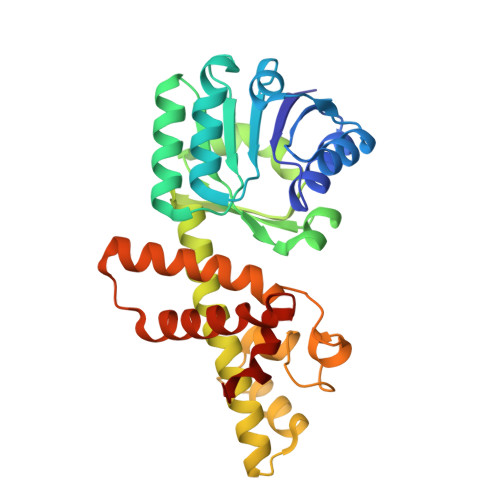
5Y8O

Mycobacterium tuberculosis 3-Hydroxyisobutyrate dehydrogenase (MtHIBADH) + NAD + 3-Hydroxy propionate (3-HP)


Experimental Data Snapshot

  • Method: X-RAY DIFFRACTION
  • Resolution: 2.05 Å
  • R-Value Free: 0.225 
  • R-Value Work: 0.176 
  • R-Value Observed: 0.178 

Starting Model: experimental
View more details

wwPDB Validation   3D Report Full Report


Ligand Structure Quality Assessment 


This is version 1.2 of the entry. See complete history


Literature

Structure, interactions and action ofMycobacterium tuberculosis3-hydroxyisobutyric acid dehydrogenase.

Srikalaivani, R.Singh, A.Vijayan, M.Surolia, A.

(2018) Biochem J 475: 2457-2471

  • DOI: https://doi.org/10.1042/BCJ20180271
  • Primary Citation of Related Structures:  
    5Y8G, 5Y8H, 5Y8I, 5Y8J, 5Y8K, 5Y8L, 5Y8M, 5Y8N, 5Y8O, 5Y8P

  • PubMed Abstract: 

    Biochemical and crystallographic studies on Mycobacterium tuberculosis 3-hydroxyisobutyric acid dehydrogenase ( Mt HIBADH), a member of the 3-hydroxyacid dehydrogenase superfamily, have been carried out. Gel filtration and blue native PAGE of Mt HIBADH show that the enzyme is a dimer. The enzyme preferentially uses NAD + as the cofactor and is specific to S -hydroxyisobutyric acid (HIBA). It can also use R -HIBA, l-serine and 3-hydroxypropanoic acid (3-HP) as substrates, but with much less efficiency. The pH optimum for activity is ∼11. Structures of the native enzyme, the holoenzyme, binary complexes with NAD + , S -HIBA, R -HIBA, l-serine and 3-HP and ternary complexes involving the substrates and NAD + have been determined. None of the already known structures of HIBADH contain a substrate molecule at the binding site. The structures reported here provide for the first time, among other things, a clear indication of the location and interactions of the substrates at the active site. They also define the entrance of the substrates to the active site region. The structures provide information on the role of specific residues at the active site and the entrance. The results obtained from crystal structures are consistent with solution studies including mutational analysis. They lead to the proposal of a plausible mechanism of the action of the enzyme.


  • Organizational Affiliation

    Molecular Biophysics Unit, Indian Institute of Science, Bangalore, India.


Macromolecules
Find similar proteins by:  (by identity cutoff)  |  3D Structure
Entity ID: 1
MoleculeChains Sequence LengthOrganismDetailsImage
Probable 3-hydroxyisobutyrate dehydrogenase
A, B
295Mycobacterium tuberculosis H37RvMutation(s): 0 
Gene Names: mmsBRv0751cMTV041.25c
EC: 1.1.1.31
UniProt
Find proteins for P9WNY5 (Mycobacterium tuberculosis (strain ATCC 25618 / H37Rv))
Explore P9WNY5 
Go to UniProtKB:  P9WNY5
Entity Groups  
Sequence Clusters30% Identity50% Identity70% Identity90% Identity95% Identity100% Identity
UniProt GroupP9WNY5
Sequence Annotations
Expand
  • Reference Sequence
Small Molecules
Ligands 5 Unique
IDChains Name / Formula / InChI Key2D Diagram3D Interactions
NAD
Query on NAD

Download Ideal Coordinates CCD File 
F [auth B]NICOTINAMIDE-ADENINE-DINUCLEOTIDE
C21 H27 N7 O14 P2
BAWFJGJZGIEFAR-NNYOXOHSSA-N
9ON
Query on 9ON

Download Ideal Coordinates CCD File 
E [auth A],
I [auth B]
(2~{S})-2-methylpentanedioic acid
C6 H10 O4
AQYCMVICBNBXNA-BYPYZUCNSA-N
GOL
Query on GOL

Download Ideal Coordinates CCD File 
D [auth A],
H [auth B]
GLYCEROL
C3 H8 O3
PEDCQBHIVMGVHV-UHFFFAOYSA-N
3OH
Query on 3OH

Download Ideal Coordinates CCD File 
G [auth B]3-HYDROXY-PROPANOIC ACID
C3 H6 O3
ALRHLSYJTWAHJZ-UHFFFAOYSA-N
AKR
Query on AKR

Download Ideal Coordinates CCD File 
C [auth A]ACRYLIC ACID
C3 H4 O2
NIXOWILDQLNWCW-UHFFFAOYSA-N
Experimental Data & Validation

Experimental Data

  • Method: X-RAY DIFFRACTION
  • Resolution: 2.05 Å
  • R-Value Free: 0.225 
  • R-Value Work: 0.176 
  • R-Value Observed: 0.178 
  • Space Group: P 65
Unit Cell:
Length ( Å )Angle ( ˚ )
a = 129.06α = 90
b = 129.06β = 90
c = 70.13γ = 120
Software Package:
Software NamePurpose
REFMACrefinement
iMOSFLMdata reduction
SCALAdata scaling
PHASERphasing

Structure Validation

View Full Validation Report


Ligand Structure Quality Assessment 


Entry History 

Deposition Data

Revision History  (Full details and data files)

  • Version 1.0: 2018-07-11
    Type: Initial release
  • Version 1.1: 2018-08-29
    Changes: Data collection, Database references
  • Version 1.2: 2023-11-22
    Changes: Data collection, Database references, Refinement description