6BT0

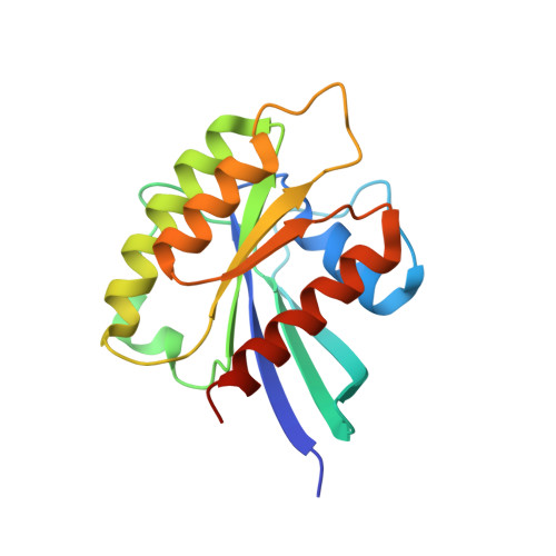
CRYSTAL STRUCTURE OF RHEB IN COMPLEX WITH COMPOUND NR1


Experimental Data Snapshot

  • Method: X-RAY DIFFRACTION
  • Resolution: 2.60 Å
  • R-Value Free: 0.316 
  • R-Value Work: 0.217 
  • R-Value Observed: 0.222 

Starting Model: experimental
View more details

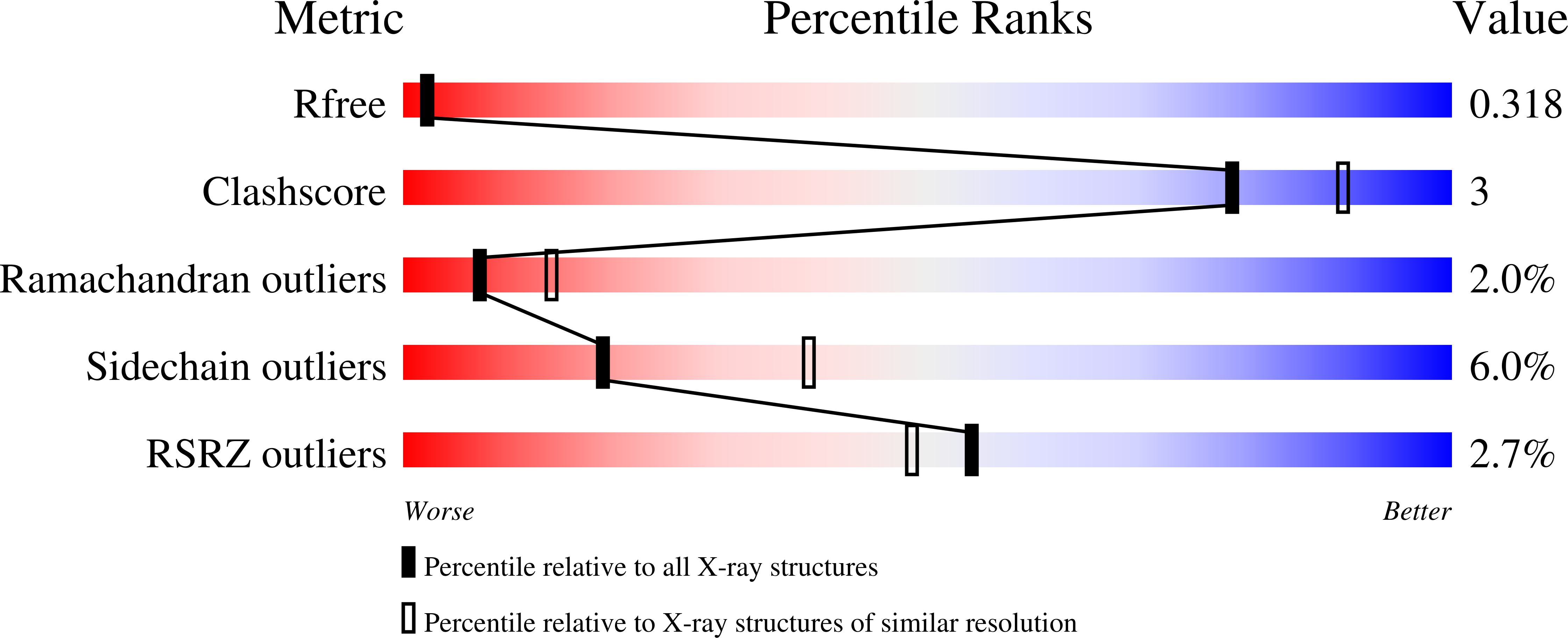
wwPDB Validation   3D Report Full Report


Ligand Structure Quality Assessment 


This is version 1.2 of the entry. See complete history


Literature

A small molecule inhibitor of Rheb selectively targets mTORC1 signaling.

Mahoney, S.J.Narayan, S.Molz, L.Berstler, L.A.Kang, S.A.Vlasuk, G.P.Saiah, E.

(2018) Nat Commun 9: 548-548

  • DOI: https://doi.org/10.1038/s41467-018-03035-z
  • Primary Citation of Related Structures:  
    5YXH, 6BSX, 6BT0

  • PubMed Abstract: 

    The small G-protein Rheb activates the mechanistic target of rapamycin complex 1 (mTORC1) in response to growth factor signals. mTORC1 is a master regulator of cellular growth and metabolism; aberrant mTORC1 signaling is associated with fibrotic, metabolic, and neurodegenerative diseases, cancers, and rare disorders. Point mutations in the Rheb switch II domain impair its ability to activate mTORC1. Here, we report the discovery of a small molecule (NR1) that binds Rheb in the switch II domain and selectively blocks mTORC1 signaling. NR1 potently inhibits mTORC1 driven phosphorylation of ribosomal protein S6 kinase beta-1 (S6K1) but does not inhibit phosphorylation of AKT or ERK. In contrast to rapamycin, NR1 does not cause inhibition of mTORC2 upon prolonged treatment. Furthermore, NR1 potently and selectively inhibits mTORC1 in mouse kidney and muscle in vivo. The data presented herein suggest that pharmacological inhibition of Rheb is an effective approach for selective inhibition of mTORC1 with therapeutic potential.


  • Organizational Affiliation

    Navitor Pharmaceuticals, Inc., 1030 Massachusetts Ave. #410, Cambridge, MA, 02138, USA. [email protected].


Macromolecules
Find similar proteins by:  (by identity cutoff)  |  3D Structure
Entity ID: 1
MoleculeChains Sequence LengthOrganismDetailsImage
GTP-binding protein Rheb
A, B, C, D
178Homo sapiensMutation(s): 0 
Gene Names: RHEBRHEB2
EC: 3.6.5
UniProt & NIH Common Fund Data Resources
Find proteins for Q15382 (Homo sapiens)
Explore Q15382 
Go to UniProtKB:  Q15382
PHAROS:  Q15382
GTEx:  ENSG00000106615 
Entity Groups  
Sequence Clusters30% Identity50% Identity70% Identity90% Identity95% Identity100% Identity
UniProt GroupQ15382
Sequence Annotations
Expand
  • Reference Sequence
Small Molecules
Ligands 3 Unique
IDChains Name / Formula / InChI Key2D Diagram3D Interactions
E7V (Subject of Investigation/LOI)
Query on E7V

Download Ideal Coordinates CCD File 
I [auth B]4-bromo-6-[(3,4-dichlorophenyl)sulfanyl]-1-{[4-(dimethylcarbamoyl)phenyl]methyl}-1H-indole-2-carboxylic acid
C25 H19 Br Cl2 N2 O3 S
MJYFVDNMTKLGTH-UHFFFAOYSA-N
GDP
Query on GDP

Download Ideal Coordinates CCD File 
F [auth A],
H [auth B],
K [auth C],
M [auth D]
GUANOSINE-5'-DIPHOSPHATE
C10 H15 N5 O11 P2
QGWNDRXFNXRZMB-UUOKFMHZSA-N
MG
Query on MG

Download Ideal Coordinates CCD File 
E [auth A],
G [auth B],
J [auth C],
L [auth D]
MAGNESIUM ION
Mg
JLVVSXFLKOJNIY-UHFFFAOYSA-N
Experimental Data & Validation

Experimental Data

  • Method: X-RAY DIFFRACTION
  • Resolution: 2.60 Å
  • R-Value Free: 0.316 
  • R-Value Work: 0.217 
  • R-Value Observed: 0.222 
  • Space Group: P 1
Unit Cell:
Length ( Å )Angle ( ˚ )
a = 49.15α = 68.35
b = 60.14β = 90.01
c = 66.32γ = 72.05
Software Package:
Software NamePurpose
XSCALEdata scaling
REFMACrefinement
PDB_EXTRACTdata extraction
XDSdata reduction
MOLREPphasing

Structure Validation

View Full Validation Report


Ligand Structure Quality Assessment 


Entry History 

Deposition Data

Revision History  (Full details and data files)

  • Version 1.0: 2018-02-28
    Type: Initial release
  • Version 1.1: 2020-10-21
    Changes: Data collection, Derived calculations
  • Version 1.2: 2023-10-04
    Changes: Data collection, Database references, Refinement description