6CBA

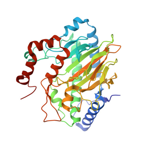
Ethylene forming enzyme in complex with manganese, 2-oxoglutarate and canavanine


Experimental Data Snapshot

  • Method: X-RAY DIFFRACTION
  • Resolution: 1.13 Å
  • R-Value Free: 0.163 
  • R-Value Work: 0.141 
  • R-Value Observed: 0.143 

Starting Model: experimental
View more details

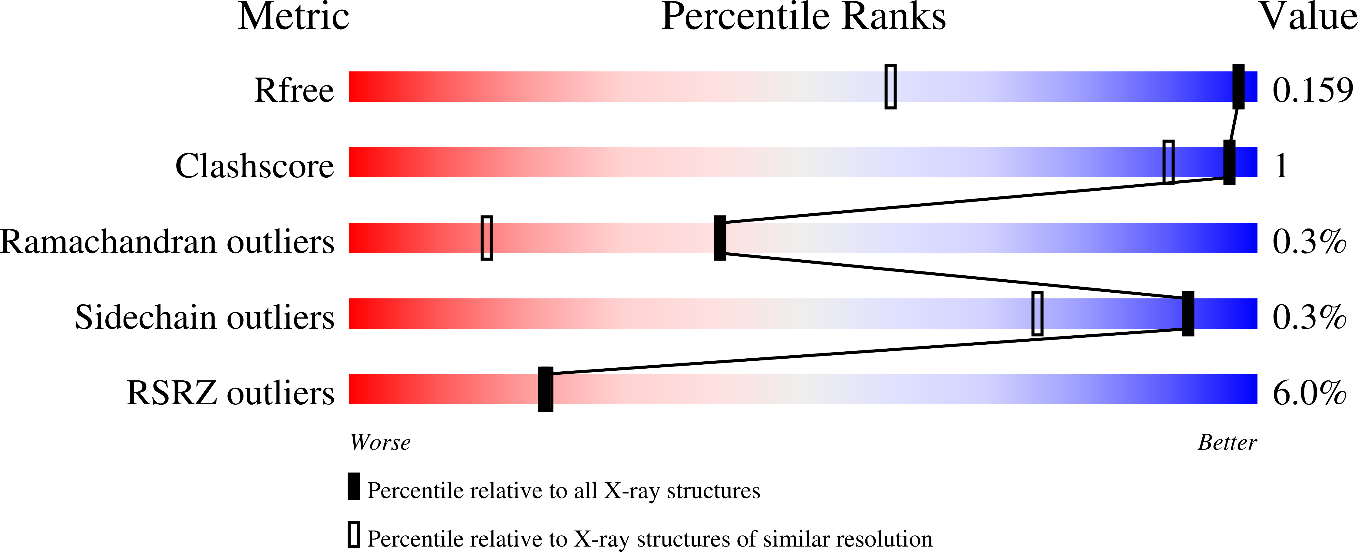
wwPDB Validation   3D Report Full Report


Ligand Structure Quality Assessment 


This is version 2.1 of the entry. See complete history


Literature

Structural, Spectroscopic, and Computational Insights from Canavanine-Bound and Two Catalytically Compromised Variants of the Ethylene-Forming Enzyme.

Chatterjee, S.Fellner, M.Rankin, J.Thomas, M.G.J S Rifayee, S.B.Christov, C.Z.Hu, J.Hausinger, R.P.

(2024) Biochemistry 

  • DOI: https://doi.org/10.1021/acs.biochem.4c00031
  • Primary Citation of Related Structures:  
    6CBA, 6CF3, 8UC2

  • PubMed Abstract: 

    The ethylene-forming enzyme (EFE) is an Fe(II), 2-oxoglutarate (2OG), and l-arginine (l-Arg)-dependent oxygenase that either forms ethylene and three CO 2 /bicarbonate from 2OG or couples the decarboxylation of 2OG to C5 hydroxylation of l-Arg. l-Arg binds with C5 toward the metal center, causing 2OG to change from monodentate to chelate metal interaction and OD1 to OD2 switch of D191 metal coordination. We applied anaerobic UV-visible spectroscopy, X-ray crystallography, and computational approaches to three EFE systems with high-resolution structures. The ineffective l-Arg analogue l-canavanine binds to the EFE with O5 pointing away from the metal center while promoting chelate formation by 2OG but fails to switch the D191 metal coordination from OD1 to OD2. Substituting alanine for R171 that interacts with 2OG and l-Arg inactivates the protein, prevents metal chelation by 2OG, and weakens l-Arg binding. The R171A EFE had electron density at the 2OG binding site that was identified by mass spectrometry as benzoic acid. The substitution by alanine of Y306 in the EFE, a residue 12 Å away from the catalytic metal center, generates an interior cavity that leads to multiple local and distal structural changes that reduce l-Arg binding and significantly reduce the enzyme activity. Flexibility analyses revealed correlated and anticorrelated motions in each system, with important distinctions from the wild-type enzyme. In combination, the results are congruent with the currently proposed enzyme mechanism, reinforce the importance of metal coordination by OD2 of D191, and highlight the importance of the second coordination sphere and longer range interactions in promoting EFE activity.


  • Organizational Affiliation

    Department of Microbiology, Genetics, and Immunology, Michigan State University, East Lansing, Michigan 48824, United States.


Macromolecules
Find similar proteins by:  (by identity cutoff)  |  3D Structure
Entity ID: 1
MoleculeChains Sequence LengthOrganismDetailsImage
2-oxoglutarate-dependent ethylene/succinate-forming enzyme352Pseudomonas savastanoi pv. phaseolicolaMutation(s): 0 
Gene Names: efe
EC: 1.13.12.19 (PDB Primary Data), 1.14.11.34 (PDB Primary Data), 1.14.20.7 (UniProt)
UniProt
Find proteins for P32021 (Pseudomonas savastanoi pv. phaseolicola)
Explore P32021 
Go to UniProtKB:  P32021
Entity Groups  
Sequence Clusters30% Identity50% Identity70% Identity90% Identity95% Identity100% Identity
UniProt GroupP32021
Sequence Annotations
Expand
  • Reference Sequence
Experimental Data & Validation

Experimental Data

  • Method: X-RAY DIFFRACTION
  • Resolution: 1.13 Å
  • R-Value Free: 0.163 
  • R-Value Work: 0.141 
  • R-Value Observed: 0.143 
  • Space Group: P 21 21 21
Unit Cell:
Length ( Å )Angle ( ˚ )
a = 48.43α = 90
b = 81.57β = 90
c = 87.63γ = 90
Software Package:
Software NamePurpose
MOSFLMdata reduction
Aimlessdata scaling
PHASERphasing
PHENIXrefinement
PDB_EXTRACTdata extraction

Structure Validation

View Full Validation Report


Ligand Structure Quality Assessment 


Entry History & Funding Information

Deposition Data


Funding OrganizationLocationGrant Number
National Institutes of Health/National Institute of General Medical Sciences (NIH/NIGMS)United StatesGM063584

Revision History  (Full details and data files)

  • Version 1.0: 2019-02-06
    Type: Initial release
  • Version 1.1: 2019-02-20
    Changes: Author supporting evidence, Data collection
  • Version 1.2: 2020-01-01
    Changes: Author supporting evidence
  • Version 1.3: 2023-10-04
    Changes: Data collection, Database references, Derived calculations, Refinement description
  • Version 2.0: 2023-11-15
    Changes: Atomic model, Data collection
  • Version 2.1: 2024-04-17
    Changes: Database references