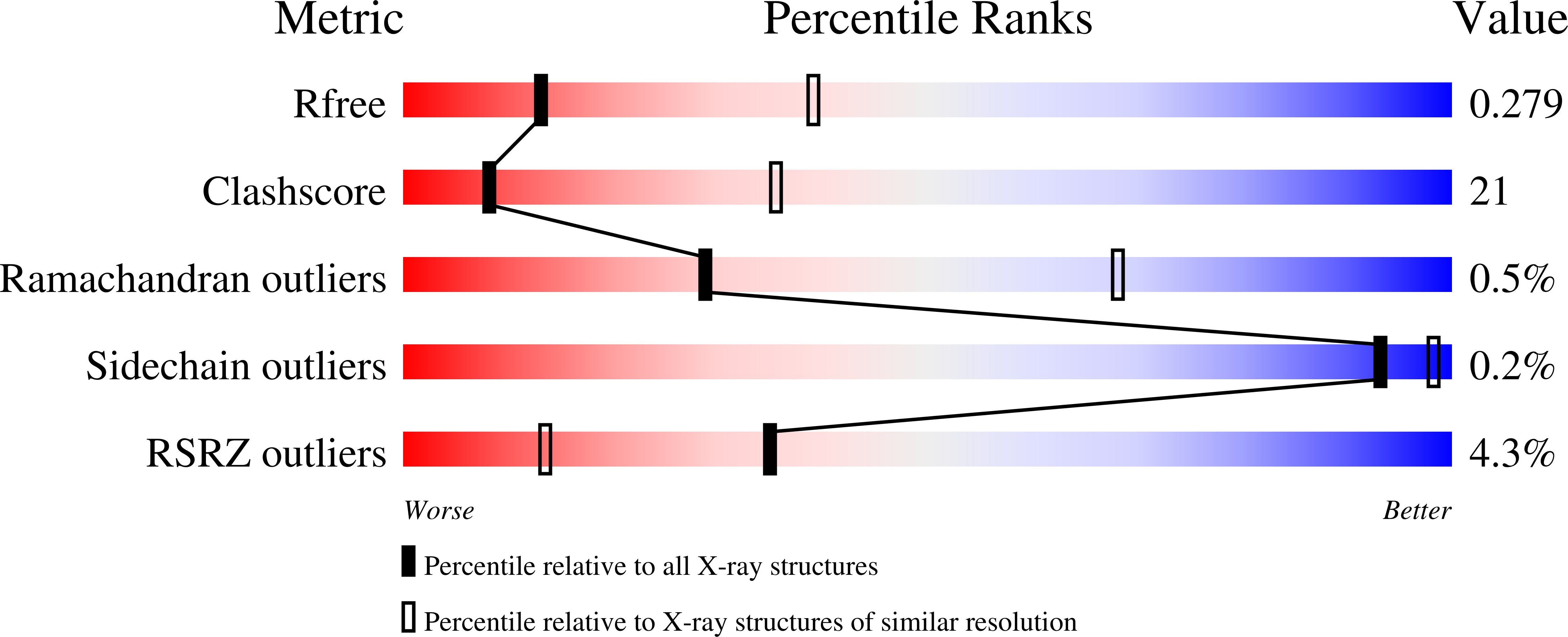
6CQ8

K2P2.1(TREK-1):ML335 complex


Experimental Data Snapshot

  • Method: X-RAY DIFFRACTION
  • Resolution: 3.00 Å
  • R-Value Free: 0.277 
  • R-Value Work: 0.257 
  • R-Value Observed: 0.258 

Starting Model: experimental
View more details

wwPDB Validation   3D Report Full Report


Ligand Structure Quality Assessment 


This is version 1.5 of the entry. See complete history


Literature

K2P2.1 (TREK-1)-activator complexes reveal a cryptic selectivity filter binding site.

Lolicato, M.Arrigoni, C.Mori, T.Sekioka, Y.Bryant, C.Clark, K.A.Minor, D.L.

(2017) Nature 547: 364-368

  • DOI: https://doi.org/10.1038/nature22988
  • Primary Citation of Related Structures:  
    6CQ6, 6CQ8, 6CQ9

  • PubMed Abstract: 

    Polymodal thermo- and mechanosensitive two-pore domain potassium (K 2P ) channels of the TREK subfamily generate 'leak' currents that regulate neuronal excitability, respond to lipids, temperature and mechanical stretch, and influence pain, temperature perception and anaesthetic responses. These dimeric voltage-gated ion channel (VGIC) superfamily members have a unique topology comprising two pore-forming regions per subunit. In contrast to other potassium channels, K 2P channels use a selectivity filter 'C-type' gate as the principal gating site. Despite recent advances, poor pharmacological profiles of K 2P channels limit mechanistic and biological studies. Here we describe a class of small-molecule TREK activators that directly stimulate the C-type gate by acting as molecular wedges that restrict interdomain interface movement behind the selectivity filter. Structures of K 2P 2.1 (also known as TREK-1) alone and with two selective K 2P 2.1 (TREK-1) and K 2P 10.1 (TREK-2) activators-an N-aryl-sulfonamide, ML335, and a thiophene-carboxamide, ML402-define a cryptic binding pocket unlike other ion channel small-molecule binding sites and, together with functional studies, identify a cation-π interaction that controls selectivity. Together, our data reveal a druggable K 2P site that stabilizes the C-type gate 'leak mode' and provide direct evidence for K 2P selectivity filter gating.


  • Organizational Affiliation

    Cardiovascular Research Institute, University of California, San Francisco, California 941158-9001, USA.


Macromolecules
Find similar proteins by:  (by identity cutoff)  |  3D Structure
Entity ID: 1
MoleculeChains Sequence LengthOrganismDetailsImage
Potassium channel subfamily K member 2
A, B
312Mus musculusMutation(s): 15 
Gene Names: Kcnk2
Membrane Entity: Yes 
UniProt & NIH Common Fund Data Resources
Find proteins for P97438 (Mus musculus)
Explore P97438 
Go to UniProtKB:  P97438
IMPC:  MGI:109366
Entity Groups  
Sequence Clusters30% Identity50% Identity70% Identity90% Identity95% Identity100% Identity
UniProt GroupP97438
Sequence Annotations
Expand
  • Reference Sequence
Small Molecules
Ligands 5 Unique
IDChains Name / Formula / InChI Key2D Diagram3D Interactions
16C
Query on 16C

Download Ideal Coordinates CCD File 
I [auth A]N-((E,2S,3R)-1,3-DIHYDROXYOCTADEC-4-EN-2-YL)PALMITAMIDE
C34 H67 N O3
YDNKGFDKKRUKPY-TURZORIXSA-N
Q6F
Query on Q6F

Download Ideal Coordinates CCD File 
H [auth A],
V [auth B]
N-[(2,4-dichlorophenyl)methyl]-4-[(methylsulfonyl)amino]benzamide
C15 H14 Cl2 N2 O3 S
RDFIQTZRJRVFHK-UHFFFAOYSA-N
R16
Query on R16

Download Ideal Coordinates CCD File 
F [auth A]
G [auth A]
J [auth A]
K [auth A]
L [auth A]
F [auth A],
G [auth A],
J [auth A],
K [auth A],
L [auth A],
S [auth B],
T [auth B],
U [auth B],
W [auth B],
X [auth B]
HEXADECANE
C16 H34
DCAYPVUWAIABOU-UHFFFAOYSA-N
CD
Query on CD

Download Ideal Coordinates CCD File 
P [auth B],
Q [auth B],
R [auth B]
CADMIUM ION
Cd
WLZRMCYVCSSEQC-UHFFFAOYSA-N
K
Query on K

Download Ideal Coordinates CCD File 
C [auth A]
D [auth A]
E [auth A]
M [auth B]
N [auth B]
C [auth A],
D [auth A],
E [auth A],
M [auth B],
N [auth B],
O [auth B]
POTASSIUM ION
K
NPYPAHLBTDXSSS-UHFFFAOYSA-N
Experimental Data & Validation

Experimental Data

  • Method: X-RAY DIFFRACTION
  • Resolution: 3.00 Å
  • R-Value Free: 0.277 
  • R-Value Work: 0.257 
  • R-Value Observed: 0.258 
  • Space Group: P 21 21 21
Unit Cell:
Length ( Å )Angle ( ˚ )
a = 67.074α = 90
b = 119.386β = 90
c = 128.179γ = 90
Software Package:
Software NamePurpose
PHENIXrefinement
Cootmodel building
Aimlessdata scaling
PHASERphasing
XDSdata processing
XDSdata reduction

Structure Validation

View Full Validation Report


Ligand Structure Quality Assessment 


Entry History & Funding Information

Deposition Data


Funding OrganizationLocationGrant Number
National Institutes of Health/National Institute of Mental Health (NIH/NIMH)United States--
American Heart AssociationUnited States--

Revision History  (Full details and data files)

  • Version 1.0: 2018-03-28
    Type: Initial release
  • Version 1.1: 2019-02-20
    Changes: Author supporting evidence, Data collection
  • Version 1.2: 2019-11-06
    Changes: Author supporting evidence, Data collection, Database references
  • Version 1.3: 2019-11-27
    Changes: Author supporting evidence
  • Version 1.4: 2023-10-04
    Changes: Data collection, Database references, Derived calculations, Refinement description, Structure summary
  • Version 1.5: 2024-10-23
    Changes: Structure summary