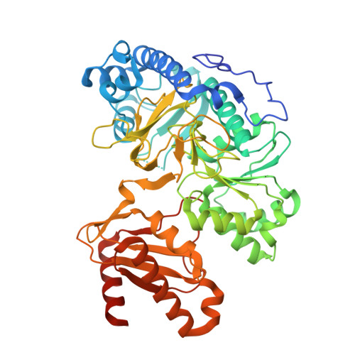
Structures of 2-Hydroxyisobutyric Acid-CoA Ligase Reveal Determinants of Substrate Specificity and Describe a Multi-Conformational Catalytic Cycle.
Zahn, M., Kurteva-Yaneva, N., Schuster, J., Krug, U., Georgi, T., Muller, R.H., Rohwerder, T., Strater, N.(2019) J Mol Biol 431: 2747-2761
- PubMed: 31145912
- DOI: https://doi.org/10.1016/j.jmb.2019.05.027
- Primary Citation of Related Structures:
6HDW, 6HDX, 6HDY, 6HE0, 6HE2 - PubMed Abstract:
2-Hydroxyisobutyric acid (2-HIBA) is a biomarker of adiposity and associated metabolic diseases such as diabetes mellitus. It is also formed in the bacterial degradation pathway of the fuel oxygenate methyl tert-butyl ether (MTBE), requiring thioesterification with CoA prior to isomerization to 3-hydroxybutyryl-CoA by B 12 -dependent acyl-CoA mutases. Here, we identify the adenylating enzymes superfamily member 2-HIBA-CoA ligase (HCL) in the MTBE-degrading bacterium Aquincola tertiaricarbonis L108 by knockout experiments. To characterize this central enzyme of 2-HIBA metabolism, ligase activity kinetics of purified HCL and its X-ray crystal structures were studied. We analyzed the enzyme in three states, which differ in the orientation of the two enzyme domains. A 154° rotation of the C-terminal domain accompanies the switch from the adenylate- into the thioester-forming state. Furthermore, a third conformation was obtained, which differs by 50° and 130° from the adenylation and thioesterification states, respectively. Phylogenetic and structural analysis reveals that HCL defines a new subgroup within phenylacetate-CoA ligases (PCLs) thus far described to exclusively accept aromatic acyl substrates. In contrast, kinetic characterization clearly demonstrated that HCL catalyzes CoA activation of several aliphatic short-chain carboxylic acids, preferentially 2-HIBA. Compared to the classical PCL representatives PaaK1 and PaaK2 of Burkholderia cenocepacia J2315, the acyl binding pocket of HCL is significantly smaller and more polar, due to unique active-site residues Y164 and S239 forming H-bonds with the OH-group of the acyl substrate moiety. Furthermore, HCL and PaaK topologies illustrate the evolutionary steps leading from a homodimeric to the fused monomeric core fold found in other ligases.
Organizational Affiliation:
Institute of Bioanalytical Chemistry, Center for Biotechnology and Biomedicine, Leipzig University, Deutscher Platz 5, 04103 Leipzig, Germany.


