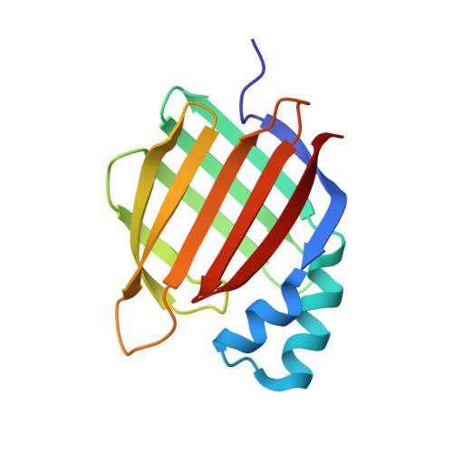
6HKR

Human Cellular Retinoic Acid Binding Protein II (CRABPII) with bound synthetic retinoid DC271.


Experimental Data Snapshot

  • Method: X-RAY DIFFRACTION
  • Resolution: 1.50 Å
  • R-Value Free: 0.175 
  • R-Value Work: 0.143 
  • R-Value Observed: 0.145 

Starting Model: experimental
View more details

wwPDB Validation   3D Report Full Report


Ligand Structure Quality Assessment 


This is version 1.2 of the entry. See complete history


Literature

Novel Fluorescence Competition Assay for Retinoic Acid Binding Proteins.

Tomlinson, C.W.E.Chisholm, D.R.Valentine, R.Whiting, A.Pohl, E.

(2018) ACS Med Chem Lett 9: 1297-1300

  • DOI: https://doi.org/10.1021/acsmedchemlett.8b00420
  • Primary Citation of Related Structures:  
    6HKR

  • PubMed Abstract: 

    Vitamin A derived retinoid compounds have multiple, powerful roles in the cellular growth and development cycle and, as a result, have attracted significant attention from both academic and pharmaceutical research in developing and characterizing synthetic retinoid analogues. Simplifying the hit development workflow for retinoid signaling will improve options available for tackling related pathologies, including tumor growth and neurodegeneration. Here, we present a novel assay that employs an intrinsically fluorescent synthetic retinoid, DC271, which allows direct measurement of the binding of nonlabeled compounds to relevant proteins. The method allows for straightforward initial measurement of binding using existing compound libraries and is followed by calculation of binding constants using a dilution series of plausible hits. The ease of use, high throughput format, and measurement of both qualitative and quantitative binding offer a new direction for retinoid-related pharmacological development.


  • Organizational Affiliation

    Department of Chemistry, Durham University, Science Laboratories, South Road, Durham, DH1 3LE, U.K.


Macromolecules
Find similar proteins by:  (by identity cutoff)  |  3D Structure
Entity ID: 1
MoleculeChains Sequence LengthOrganismDetailsImage
Cellular retinoic acid-binding protein 2138Homo sapiensMutation(s): 0 
Gene Names: CRABP2
UniProt & NIH Common Fund Data Resources
Find proteins for P29373 (Homo sapiens)
Explore P29373 
Go to UniProtKB:  P29373
PHAROS:  P29373
GTEx:  ENSG00000143320 
Entity Groups  
Sequence Clusters30% Identity50% Identity70% Identity90% Identity95% Identity100% Identity
UniProt GroupP29373
Sequence Annotations
Expand
  • Reference Sequence
Small Molecules
Ligands 4 Unique
IDChains Name / Formula / InChI Key2D Diagram3D Interactions
G9Q (Subject of Investigation/LOI)
Query on G9Q

Download Ideal Coordinates CCD File 
C [auth A]4-[2-(4,4-dimethyl-1-propan-2-yl-2,3-dihydroquinolin-6-yl)ethynyl]benzoic acid
C23 H25 N O2
OCMSZODRCJAGHL-UHFFFAOYSA-N
PG4
Query on PG4

Download Ideal Coordinates CCD File 
F [auth A]TETRAETHYLENE GLYCOL
C8 H18 O5
UWHCKJMYHZGTIT-UHFFFAOYSA-N
GOL
Query on GOL

Download Ideal Coordinates CCD File 
D [auth A],
E [auth A]
GLYCEROL
C3 H8 O3
PEDCQBHIVMGVHV-UHFFFAOYSA-N
EDO
Query on EDO

Download Ideal Coordinates CCD File 
B [auth A]1,2-ETHANEDIOL
C2 H6 O2
LYCAIKOWRPUZTN-UHFFFAOYSA-N
Experimental Data & Validation

Experimental Data

  • Method: X-RAY DIFFRACTION
  • Resolution: 1.50 Å
  • R-Value Free: 0.175 
  • R-Value Work: 0.143 
  • R-Value Observed: 0.145 
  • Space Group: P 21 21 21
Unit Cell:
Length ( Å )Angle ( ˚ )
a = 44.689α = 90
b = 47.479β = 90
c = 77.733γ = 90
Software Package:
Software NamePurpose
REFMACrefinement
xia2data reduction
xia2data scaling
PHASERphasing

Structure Validation

View Full Validation Report


Ligand Structure Quality Assessment 


Entry History & Funding Information

Deposition Data


Funding OrganizationLocationGrant Number
Medical Research Council (United Kingdom)United Kingdom--

Revision History  (Full details and data files)

  • Version 1.0: 2018-11-28
    Type: Initial release
  • Version 1.1: 2019-01-16
    Changes: Data collection, Database references
  • Version 1.2: 2024-01-17
    Changes: Data collection, Database references, Refinement description