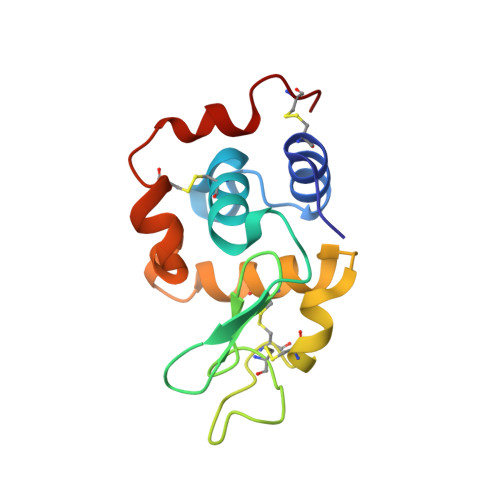
6IG6

Crystal structure of lysozyme delivered in polyacrylamide using x-ray free electron laser

  • Classification: HYDROLASE
  • Organism(s): Gallus gallus
  • Expression System: Gallus gallus
  • Mutation(s): No 

  • Deposited: 2018-09-25 Released: 2018-11-07 
  • Deposition Author(s): Nam, K.H.
  • Funding Organization(s): National Research Foundation (Korea)

Experimental Data Snapshot

  • Method: X-RAY DIFFRACTION
  • Resolution: 1.70 Å
  • R-Value Free: 0.217 
  • R-Value Work: 0.196 
  • R-Value Observed: 0.198 

Starting Model: experimental
View more details

wwPDB Validation   3D Report Full Report


This is version 1.7 of the entry. See complete history


Literature

Polyacrylamide injection matrix for serial femtosecond crystallography.

Park, J.Park, S.Kim, J.Park, G.Cho, Y.Nam, K.H.

(2019) Sci Rep 9: 2525-2525

  • DOI: https://doi.org/10.1038/s41598-019-39020-9
  • Primary Citation of Related Structures:  
    6IG6, 6IG7

  • PubMed Abstract: 

    Serial femtosecond crystallography (SFX) provides opportunities to observe the dynamics of macromolecules without causing radiation damage at room temperature. Although SFX provides a biologically more reliable crystal structure than provided by the existing synchrotron sources, there are limitations due to the consumption of many crystal samples. A viscous medium as a carrier matrix reduces the flow rate of the crystal sample from the injector, thereby dramatically reducing sample consumption. However, the currently available media cannot be applied to specific crystal samples owing to reactions between the viscous medium and crystal sample. The discovery and characterisation of a new delivery medium for SFX can further expand its use. Herein, we report the preparation of a polyacrylamide (PAM) injection matrix to determine the crystal structure with an X-ray free-electron laser. We obtained 11,936 and 22,213 indexed images using 0.5 mg lysozyme and 1.0 mg thermolysin, respectively. We determined the crystal structures of lysozyme and thermolysin delivered in PAM at 1.7 Å and 1.8 Å resolutions. The maximum background scattering from PAM was lower than monoolein, a commonly used viscous medium. Our results show that PAM can be used as a sample delivery media in SFX studies.


  • Organizational Affiliation

    Pohang Accelerator Laboratory, Pohang, Gyeongbuk, Republic of Korea. [email protected].


Macromolecules
Find similar proteins by:  (by identity cutoff)  |  3D Structure
Entity ID: 1
MoleculeChains Sequence LengthOrganismDetailsImage
Lysozyme C129Gallus gallusMutation(s): 0 
Gene Names: LYZ
EC: 3.2.1.17
UniProt
Find proteins for P00698 (Gallus gallus)
Explore P00698 
Go to UniProtKB:  P00698
Entity Groups  
Sequence Clusters30% Identity50% Identity70% Identity90% Identity95% Identity100% Identity
UniProt GroupP00698
Sequence Annotations
Expand
  • Reference Sequence
Experimental Data & Validation

Experimental Data

Unit Cell:
Length ( Å )Angle ( ˚ )
a = 79.6α = 90
b = 79.6β = 90
c = 38.2γ = 90
Software Package:
Software NamePurpose
PHASERphasing
PHENIXrefinement
PDB_EXTRACTdata extraction
CrystFELdata reduction
CrystFELdata scaling

Structure Validation

View Full Validation Report


Entry History & Funding Information

Deposition Data

  • Released Date: 2018-11-07 
  • Deposition Author(s): Nam, K.H.

Funding OrganizationLocationGrant Number
National Research Foundation (Korea)Korea, Republic OfNRF-2017M3A9F6029736

Revision History  (Full details and data files)

  • Version 1.0: 2018-11-07
    Type: Initial release
  • Version 1.1: 2019-01-23
    Changes: Data collection
  • Version 1.2: 2019-05-22
    Changes: Data collection, Database references
  • Version 1.3: 2019-08-28
    Changes: Data collection
  • Version 1.4: 2019-09-18
    Changes: Data collection
  • Version 1.5: 2023-09-06
    Changes: Data collection, Database references
  • Version 1.6: 2023-11-22
    Changes: Refinement description
  • Version 1.7: 2024-10-16
    Changes: Structure summary