6M7O

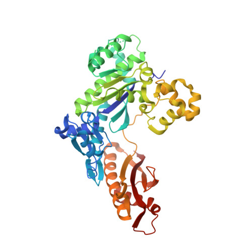
Human DNA polymerase eta ternary complex with Mn2+ and dTMPNPP oppositing cdA

  • Classification: TRANSFERASE/DNA
  • Organism(s): Homo sapiens
  • Expression System: Escherichia coli
  • Mutation(s): No 

  • Deposited: 2018-08-20 Released: 2018-09-12 
  • Deposition Author(s): Weng, P., Gao, Y., Yang, W.
  • Funding Organization(s): National Institutes of Health/National Institute of Diabetes and Digestive and Kidney Disease (NIH/NIDDK)

Experimental Data Snapshot

  • Method: X-RAY DIFFRACTION
  • Resolution: 3.00 Å
  • R-Value Free: 0.273 
  • R-Value Work: 0.231 
  • R-Value Observed: 0.233 

Starting Model: experimental
View more details

wwPDB Validation   3D Report Full Report


Ligand Structure Quality Assessment 


This is version 1.4 of the entry. See complete history


Literature

Bypassing a 8,5'-cyclo-2'-deoxyadenosine lesion by human DNA polymerase eta at atomic resolution.

Weng, P.J.Gao, Y.Gregory, M.T.Wang, P.Wang, Y.Yang, W.

(2018) Proc Natl Acad Sci U S A 115: 10660-10665

  • DOI: https://doi.org/10.1073/pnas.1812856115
  • Primary Citation of Related Structures:  
    6M7O, 6M7P, 6M7T, 6M7U, 6M7V

  • PubMed Abstract: 

    Oxidatively induced DNA lesions 8,5'-cyclopurine-2'-deoxynucleosides (cdPus) are prevalent and cytotoxic by impeding DNA replication and transcription. Both the 5' R - and 5' S -diastereomers of cdPu can be removed by nucleotide excision repair; however, the 5' S -cdPu is more resistant to repair than the 5' R counterpart. Here, we report the crystal structures of human polymerase (Pol) η bypassing 5' S -8,5'-cyclo-2'-deoxyadenosine (cdA) in insertion and the following two extension steps. The cdA-containing DNA structures vary in response to the protein environment. Supported by the "molecular splint" of Pol η, the structure of 5' S -cdA at 1.75-Å resolution reveals that the backbone is pinched toward the minor groove and the adenine base is tilted. In the templating position, the cdA takes up the extra space usually reserved for the thymine dimer, and dTTP is efficiently incorporated by Pol η in the presence of Mn 2+ Rigid distortions of the DNA duplex by cdA, however, prevent normal base pairing and hinder immediate primer extension by Pol η. Our results provide structural insights into the strong replication blockage effect and the mutagenic property of the cdPu lesions in cells.


  • Organizational Affiliation

    Laboratory of Molecular Biology, National Institute of Diabetes and Digestive and Kidney Diseases, National Institutes of Health, Bethesda, MD 20892.


Macromolecules

Find similar proteins by:  (by identity cutoff)  |  3D Structure
Entity ID: 1
MoleculeChains Sequence LengthOrganismDetailsImage
DNA polymerase eta435Homo sapiensMutation(s): 0 
Gene Names: POLHRAD30RAD30AXPV
EC: 2.7.7.7
UniProt & NIH Common Fund Data Resources
Find proteins for Q9Y253 (Homo sapiens)
Explore Q9Y253 
Go to UniProtKB:  Q9Y253
PHAROS:  Q9Y253
GTEx:  ENSG00000170734 
Entity Groups  
Sequence Clusters30% Identity50% Identity70% Identity90% Identity95% Identity100% Identity
UniProt GroupQ9Y253
Sequence Annotations
Expand
  • Reference Sequence

Find similar nucleic acids by:  Sequence   |   3D Structure  

Entity ID: 2
MoleculeChains LengthOrganismImage
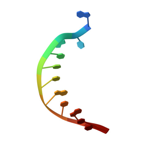
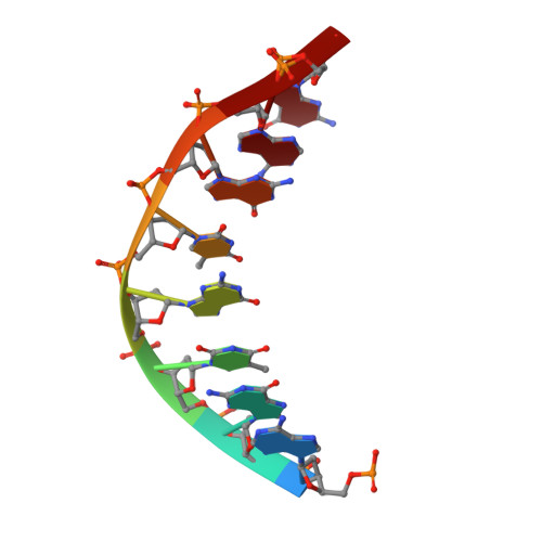
DNA (5'-D(*AP*TP*(02I)P*CP*TP*CP*AP*CP*AP*CP*T)-3')B [auth T]11Homo sapiens
Sequence Annotations
Expand
  • Reference Sequence

Find similar nucleic acids by:  Sequence   |   3D Structure  

Entity ID: 3
MoleculeChains LengthOrganismImage
DNA (5'-D(P*AP*GP*TP*GP*TP*GP*AP*G*(1FZ))-3')C [auth P]8Homo sapiens
Sequence Annotations
Expand
  • Reference Sequence
Experimental Data & Validation

Experimental Data

  • Method: X-RAY DIFFRACTION
  • Resolution: 3.00 Å
  • R-Value Free: 0.273 
  • R-Value Work: 0.231 
  • R-Value Observed: 0.233 
  • Space Group: P 61
Unit Cell:
Length ( Å )Angle ( ˚ )
a = 98.9α = 90
b = 98.9β = 90
c = 81.06γ = 120
Software Package:
Software NamePurpose
PHENIXrefinement
XDSdata reduction
XSCALEdata scaling
PHENIXphasing

Structure Validation

View Full Validation Report


Ligand Structure Quality Assessment 


Entry History & Funding Information

Deposition Data


Funding OrganizationLocationGrant Number
National Institutes of Health/National Institute of Diabetes and Digestive and Kidney Disease (NIH/NIDDK)United StatesDK036146

Revision History  (Full details and data files)

  • Version 1.0: 2018-09-12
    Type: Initial release
  • Version 1.1: 2018-10-17
    Changes: Data collection, Database references
  • Version 1.2: 2018-10-31
    Changes: Data collection, Database references
  • Version 1.3: 2019-12-25
    Changes: Author supporting evidence
  • Version 1.4: 2023-10-11
    Changes: Data collection, Database references, Derived calculations, Refinement description