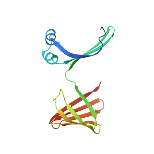
6MCV

Crystal Structure of Holo Retinal-Bound Domain-Swapped Dimer of Wild Type Human Cellular Retinol Binding Protein II


Experimental Data Snapshot

  • Method: X-RAY DIFFRACTION
  • Resolution: 3.30 Å
  • R-Value Free: 0.266 
  • R-Value Work: 0.208 
  • R-Value Observed: 0.210 

Starting Model: experimental
View more details

wwPDB Validation   3D Report Full Report


Ligand Structure Quality Assessment 


This is version 1.3 of the entry. See complete history


Literature

Engineering the hCRBPII Domain-Swapped Dimer into a New Class of Protein Switches.

Ghanbarpour, A.Pinger, C.Esmatpour Salmani, R.Assar, Z.Santos, E.M.Nosrati, M.Pawlowski, K.Spence, D.Vasileiou, C.Jin, X.Borhan, B.Geiger, J.H.

(2019) J Am Chem Soc 141: 17125-17132

  • DOI: https://doi.org/10.1021/jacs.9b04664
  • Primary Citation of Related Structures:  
    6E50, 6E51, 6E5E, 6E5Q, 6E5R, 6E5S, 6E6L, 6E7M, 6MCU, 6MCV, 6MKV, 6MLB, 6ON5, 6ON7, 6ON8

  • PubMed Abstract: 

    Protein conformational switches or allosteric proteins play a key role in the regulation of many essential biological pathways. Nonetheless, the implementation of protein conformational switches in protein design applications has proven challenging, with only a few known examples that are not derivatives of naturally occurring allosteric systems. We have discovered that the domain-swapped (DS) dimer of hCRBPII undergoes a large and robust conformational change upon retinal binding, making it a potentially powerful template for the design of protein conformational switches. Atomic resolution structures of the apo- and holo-forms illuminate a simple, mechanical movement involving sterically driven torsion angle flipping of two residues that drive the motion. We further demonstrate that the conformational "readout" can be altered by addition of cross-domain disulfide bonds, also visualized at atomic resolution. Finally, as a proof of principle, we have created an allosteric metal binding site in the DS dimer, where ligand binding results in a reversible 5-fold loss of metal binding affinity. The high resolution structure of the metal-bound variant illustrates a well-formed metal binding site at the interface of the two domains of the DS dimer and confirms the design strategy for allosteric regulation.


  • Organizational Affiliation

    Department of Chemistry , Michigan State University , East Lansing , Michigan 48824 , United States.


Macromolecules
Find similar proteins by:  (by identity cutoff)  |  3D Structure
Entity ID: 1
MoleculeChains Sequence LengthOrganismDetailsImage
Retinol-binding protein 2
A, B, C, D, E
A, B, C, D, E, F, G, H, I, J, K, L
133Homo sapiensMutation(s): 0 
Gene Names: RBP2CRBP2
UniProt & NIH Common Fund Data Resources
Find proteins for P50120 (Homo sapiens)
Explore P50120 
Go to UniProtKB:  P50120
PHAROS:  P50120
GTEx:  ENSG00000114113 
Entity Groups  
Sequence Clusters30% Identity50% Identity70% Identity90% Identity95% Identity100% Identity
UniProt GroupP50120
Sequence Annotations
Expand
  • Reference Sequence
Experimental Data & Validation

Experimental Data

  • Method: X-RAY DIFFRACTION
  • Resolution: 3.30 Å
  • R-Value Free: 0.266 
  • R-Value Work: 0.208 
  • R-Value Observed: 0.210 
  • Space Group: P 21 21 21
Unit Cell:
Length ( Å )Angle ( ˚ )
a = 65.819α = 90
b = 74.362β = 90
c = 352.547γ = 90
Software Package:
Software NamePurpose
PHENIXrefinement
HKL-2000data reduction
HKL-2000data scaling
PHASERphasing

Structure Validation

View Full Validation Report


Ligand Structure Quality Assessment 


Entry History & Funding Information

Deposition Data


Funding OrganizationLocationGrant Number
National Institutes of Health/National Human Genome Research Institute (NIH/NHGRI)United States--

Revision History  (Full details and data files)

  • Version 1.0: 2019-10-16
    Type: Initial release
  • Version 1.1: 2019-11-13
    Changes: Data collection, Database references
  • Version 1.2: 2019-12-18
    Changes: Author supporting evidence
  • Version 1.3: 2023-10-11
    Changes: Data collection, Database references, Refinement description