6MLH

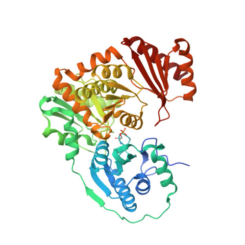
Crystal structure of X. citri phosphoglucomutase in complex with GLUCOPYRANOSYL-1-METHYL-PHOSPHONIC ACID


Experimental Data Snapshot

  • Method: X-RAY DIFFRACTION
  • Resolution: 1.65 Å
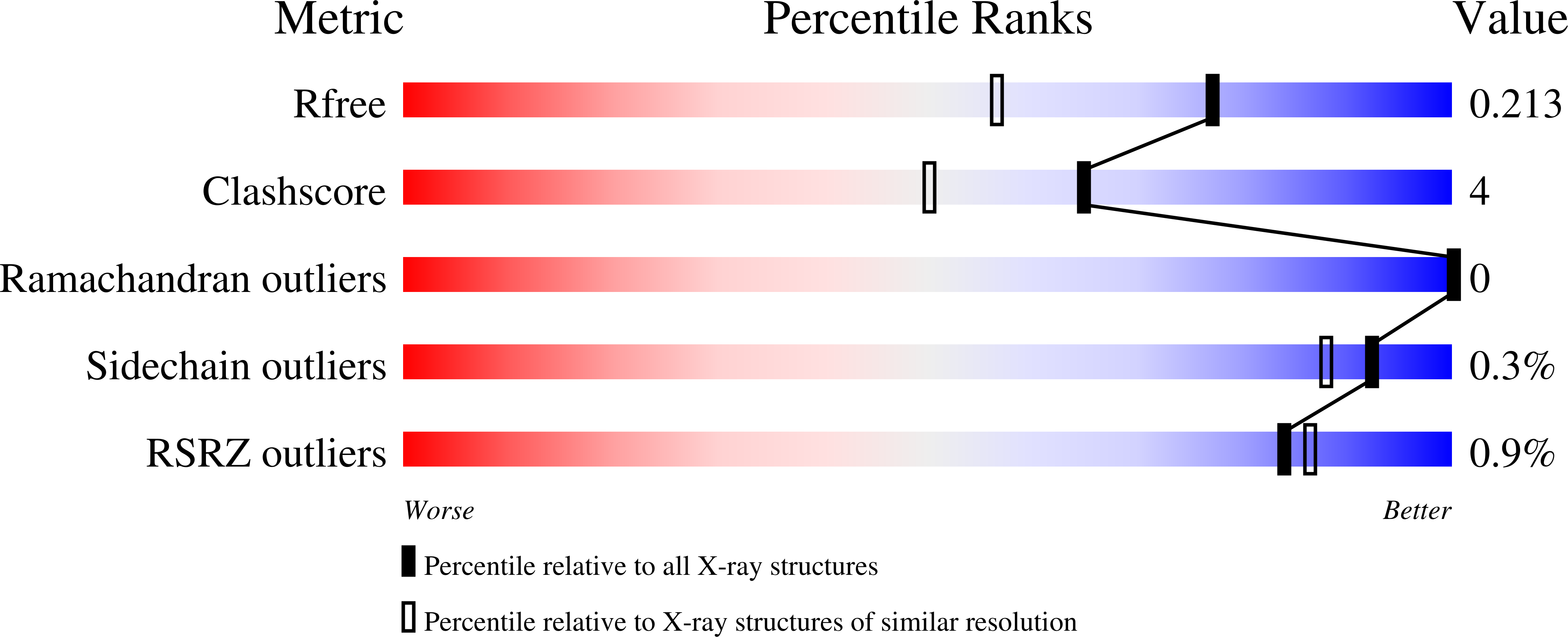
  • R-Value Free: 0.213 
  • R-Value Work: 0.172 
  • R-Value Observed: 0.174 

Starting Model: experimental
View more details

wwPDB Validation   3D Report Full Report


Ligand Structure Quality Assessment 


This is version 1.5 of the entry. See complete history


Literature

Inhibitory Evaluation of alpha PMM/PGM fromPseudomonas aeruginosa: Chemical Synthesis, Enzyme Kinetics, and Protein Crystallographic Study.

Zhu, J.S.Stiers, K.M.Soleimani, E.Groves, B.R.Beamer, L.J.Jakeman, D.L.

(2019) J Org Chem 84: 9627-9636

  • DOI: https://doi.org/10.1021/acs.joc.9b01305
  • Primary Citation of Related Structures:  
    6MLF, 6MLH, 6MLW, 6MNV

  • PubMed Abstract: 

    α-Phosphomannomutase/phosphoglucomutase (αPMM/PGM) from P. aeruginosa is involved in bacterial cell wall assembly and is implicated in P. aeruginosa virulence, yet few studies have addressed αPMM/PGM inhibition from this important Gram-negative bacterial human pathogen. Four structurally different α-d-glucopyranose 1-phosphate (αG1P) derivatives including 1- C -fluoromethylated analogues ( 1 - 3 ), 1,2-cyclic phosph(on)ate analogues ( 4 - 6 ), isosteric methylene phosphono analogues ( 7 and 8 ), and 6-fluoro-αG1P ( 9 ), were synthesized and assessed as potential time-dependent or reversible αPMM/PGM inhibitors. The resulting kinetic data were consistent with the crystallographic structures of the highly homologous Xanthomonas citri αPGM with inhibitors 3 and 7 - 9 binding to the enzyme active site (1.65-1.9 Å). These structural and kinetic insights will enhance the design of future αPMM/PGM inhibitors.


  • Organizational Affiliation

    College of Pharmacy , Dalhousie University , 5968 College Street , Halifax , Nova Scotia B3H 4R2 , Canada.


Macromolecules
Find similar proteins by:  (by identity cutoff)  |  3D Structure
Entity ID: 1
MoleculeChains Sequence LengthOrganismDetailsImage
Phosphoglucomutase468Xanthomonas citriMutation(s): 0 
Gene Names: xanA
Entity Groups  
Sequence Clusters30% Identity50% Identity70% Identity90% Identity95% Identity100% Identity
Sequence Annotations
Expand
  • Reference Sequence
Small Molecules
Modified Residues  1 Unique
IDChains TypeFormula2D DiagramParent
SEP
Query on SEP
A
L-PEPTIDE LINKINGC3 H8 N O6 PSER
Experimental Data & Validation

Experimental Data

  • Method: X-RAY DIFFRACTION
  • Resolution: 1.65 Å
  • R-Value Free: 0.213 
  • R-Value Work: 0.172 
  • R-Value Observed: 0.174 
  • Space Group: P 21 21 21
Unit Cell:
Length ( Å )Angle ( ˚ )
a = 43.571α = 90
b = 54.715β = 90
c = 172.682γ = 90
Software Package:
Software NamePurpose
PHENIXrefinement
XDSdata reduction
XDSdata scaling
PHENIXphasing

Structure Validation

View Full Validation Report


Ligand Structure Quality Assessment 


Entry History & Funding Information

Deposition Data


Funding OrganizationLocationGrant Number
National Science Foundation (NSF, United States)United StatesMCB 1409898

Revision History  (Full details and data files)

  • Version 1.0: 2019-07-31
    Type: Initial release
  • Version 1.1: 2019-08-14
    Changes: Data collection, Database references
  • Version 1.2: 2019-11-27
    Changes: Author supporting evidence
  • Version 1.3: 2020-07-29
    Type: Remediation
    Reason: Carbohydrate remediation
    Changes: Derived calculations, Structure summary
  • Version 1.4: 2023-10-11
    Changes: Data collection, Database references, Refinement description, Structure summary
  • Version 1.5: 2024-11-06
    Changes: Structure summary