6MPZ

Crystal structure of a double glycine motif protease from AMS/PCAT transporter in complex with the leader peptide


Experimental Data Snapshot

  • Method: X-RAY DIFFRACTION
  • Resolution: 2.00 Å
  • R-Value Free: 0.268 
  • R-Value Work: 0.234 
  • R-Value Observed: 0.236 

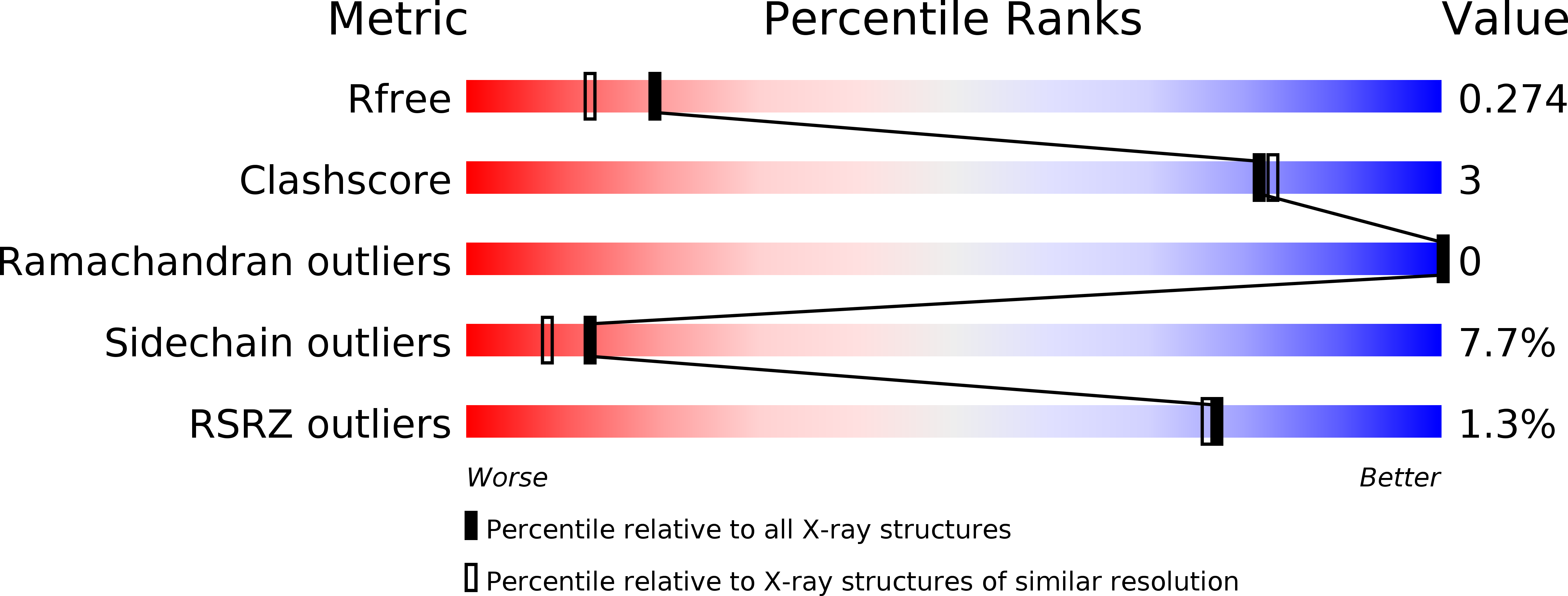
wwPDB Validation   3D Report Full Report


Ligand Structure Quality Assessment 


This is version 1.1 of the entry. See complete history


Literature

Insights into AMS/PCAT transporters from biochemical and structural characterization of a double Glycine motif protease.

Bobeica, S.C.Dong, S.Huo, L.Mazo, N.McLaughlin, M.I.H.Jimenez-Oses, G.Nair, S.K.van der Donk, W.A.

(2019) Elife 8

  • DOI: https://doi.org/10.7554/eLife.42305
  • Primary Citation of Related Structures:  
    6MPZ

  • PubMed Abstract: 

    The secretion of peptides and proteins is essential for survival and ecological adaptation of bacteria. Dual-functional ATP-binding cassette transporters export antimicrobial or quorum signaling peptides in Gram-positive bacteria. Their substrates contain a leader sequence that is excised by an N-terminal peptidase C39 domain at a double Gly motif. We characterized the protease domain (LahT150) of a transporter from a lanthipeptide biosynthetic operon in Lachnospiraceae and demonstrate that this protease can remove the leader peptide from a diverse set of peptides. The 2.0 Å resolution crystal structure of the protease domain in complex with a covalently bound leader peptide demonstrates the basis for substrate recognition across the entire class of such transporters. The structural data also provide a model for understanding the role of leader peptide recognition in the translocation cycle, and the function of degenerate, non-functional C39-like domains (CLD) in substrate recruitment in toxin exporters in Gram-negative bacteria.


  • Organizational Affiliation

    Roger Adams Laboratory, Department of Chemistry, University of Illinois at Urbana-Champaign, Urbana, United States.


Macromolecules
Find similar proteins by:  (by identity cutoff)  |  3D Structure
Entity ID: 1
MoleculeChains Sequence LengthOrganismDetailsImage
Double Glycine Motif Protease domain from AMS/PCAT Transporter
A, B, C, D
147Lachnospiraceae bacterium C6A11Mutation(s): 0 
Entity Groups  
Sequence Clusters30% Identity50% Identity70% Identity90% Identity95% Identity100% Identity
Sequence Annotations
Expand
  • Reference Sequence

Find similar proteins by:  Sequence   |   3D Structure  

Entity ID: 2
MoleculeChains Sequence LengthOrganismDetailsImage
peptide aldehyde inhibitor 1 based on the ProcA2.8 leader peptideE [auth M],
F [auth N],
G [auth O],
H [auth P]
14Prochlorococcus marinus str. MIT 9313Mutation(s): 0 
Entity Groups  
Sequence Clusters30% Identity50% Identity70% Identity90% Identity95% Identity100% Identity
Sequence Annotations
Expand
  • Reference Sequence
Small Molecules
Modified Residues  1 Unique
IDChains TypeFormula2D DiagramParent
GLZ
Query on GLZ
E [auth M],
F [auth N],
G [auth O],
H [auth P]
L-PEPTIDE LINKINGC2 H5 N OGLY
Experimental Data & Validation

Experimental Data

  • Method: X-RAY DIFFRACTION
  • Resolution: 2.00 Å
  • R-Value Free: 0.268 
  • R-Value Work: 0.234 
  • R-Value Observed: 0.236 
  • Space Group: P 1 21 1
Unit Cell:
Length ( Å )Angle ( ˚ )
a = 37.888α = 90
b = 119.426β = 93.84
c = 76.522γ = 90
Software Package:
Software NamePurpose
REFMACrefinement
XDSdata scaling
AutoSolphasing
PHENIXmodel building

Structure Validation

View Full Validation Report


Ligand Structure Quality Assessment 


Entry History & Funding Information

Deposition Data


Funding OrganizationLocationGrant Number
National Institutes of Health/National Institute of General Medical Sciences (NIH/NIGMS)United StatesGM079038

Revision History  (Full details and data files)

  • Version 1.0: 2019-02-06
    Type: Initial release
  • Version 1.1: 2020-01-01
    Changes: Author supporting evidence