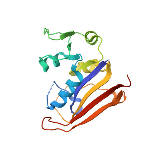
6MT8

E. coli DHFR complex modeled with two ligand states


Experimental Data Snapshot

  • Method: X-RAY DIFFRACTION
  • Resolution: 1.35 Å
  • R-Value Free: 0.190 
  • R-Value Work: 0.167 
  • R-Value Observed: 0.169 

Starting Model: experimental
View more details

wwPDB Validation   3D Report Full Report


Ligand Structure Quality Assessment 


This is version 1.2 of the entry. See complete history


Literature

Time-resolved x-ray crystallography capture of a slow reaction tetrahydrofolate intermediate.

Cao, H.Skolnick, J.

(2019) Struct Dyn 6: 024701-024701

  • DOI: https://doi.org/10.1063/1.5086436
  • Primary Citation of Related Structures:  
    6MR9, 6MT8, 6MTH

  • PubMed Abstract: 

    Time-resolved crystallography is a powerful technique to elucidate molecular mechanisms at both spatial (angstroms) and temporal (picoseconds to seconds) resolutions. We recently discovered an unusually slow reaction at room temperature that occurs on the order of days: the in crystalline reverse oxidative decay of the chemically labile (6S)-5,6,7,8-tetrahydrofolate in complex with its producing enzyme Escherichia coli dihydrofolate reductase. Here, we report the critical analysis of a representative dataset at an intermediate reaction time point. A quinonoid-like intermediate state lying between tetrahydrofolate and dihydrofolate features a near coplanar geometry of the bicyclic pterin moiety, and a tetrahedral sp 3 C6 geometry is proposed based on the apparent mFo-DFc omit electron densities of the ligand. The presence of this intermediate is strongly supported by Bayesian difference refinement. Isomorphous Fo-Fo difference map and multi-state refinement analyses suggest the presence of end-state ligand populations as well, although the putative intermediate state is likely the most populated. A similar quinonoid intermediate previously proposed to transiently exist during the oxidation of tetrahydrofolate was confirmed by polarography and UV-vis spectroscopy to be relatively stable in the oxidation of its close analog tetrahydropterin. We postulate that the constraints on the ligand imposed by the interactions with the protein environment might be the origin of the slow reaction observed by time-resolved crystallography.


  • Organizational Affiliation

    Center for the Study of Systems Biology, School of Biological Sciences, Georgia Institute of Technology, 950 Atlantic Drive, NW, Atlanta, Georgia 30332, USA.


Macromolecules
Find similar proteins by:  (by identity cutoff)  |  3D Structure
Entity ID: 1
MoleculeChains Sequence LengthOrganismDetailsImage
Dihydrofolate reductase165Escherichia coli K-12Mutation(s): 0 
Gene Names: folAtmrAb0048JW0047
EC: 1.5.1.3
UniProt
Find proteins for P0ABQ4 (Escherichia coli (strain K12))
Explore P0ABQ4 
Go to UniProtKB:  P0ABQ4
Entity Groups  
Sequence Clusters30% Identity50% Identity70% Identity90% Identity95% Identity100% Identity
UniProt GroupP0ABQ4
Sequence Annotations
Expand
  • Reference Sequence
Small Molecules
Ligands 4 Unique
IDChains Name / Formula / InChI Key2D Diagram3D Interactions
THG
Query on THG

Download Ideal Coordinates CCD File 
I [auth A](6S)-5,6,7,8-TETRAHYDROFOLATE
C19 H23 N7 O6
MSTNYGQPCMXVAQ-RYUDHWBXSA-N
DHF
Query on DHF

Download Ideal Coordinates CCD File 
J [auth A]DIHYDROFOLIC ACID
C19 H21 N7 O6
OZRNSSUDZOLUSN-LBPRGKRZSA-N
CL
Query on CL

Download Ideal Coordinates CCD File 
B [auth A],
C [auth A],
D [auth A]
CHLORIDE ION
Cl
VEXZGXHMUGYJMC-UHFFFAOYSA-M
MG
Query on MG

Download Ideal Coordinates CCD File 
E [auth A],
F [auth A],
G [auth A],
H [auth A]
MAGNESIUM ION
Mg
JLVVSXFLKOJNIY-UHFFFAOYSA-N
Experimental Data & Validation

Experimental Data

  • Method: X-RAY DIFFRACTION
  • Resolution: 1.35 Å
  • R-Value Free: 0.190 
  • R-Value Work: 0.167 
  • R-Value Observed: 0.169 
  • Space Group: P 21 21 21
Unit Cell:
Length ( Å )Angle ( ˚ )
a = 34.05α = 90
b = 51.666β = 90
c = 79.014γ = 90
Software Package:
Software NamePurpose
XDSdata reduction
XSCALEdata scaling
PHASERphasing
PHENIXrefinement
PDB_EXTRACTdata extraction

Structure Validation

View Full Validation Report


Ligand Structure Quality Assessment 


Entry History & Funding Information

Deposition Data


Funding OrganizationLocationGrant Number
National Institutes of Health/National Institute of General Medical Sciences (NIH/NIGMS)United StatesR35GM118039

Revision History  (Full details and data files)

  • Version 1.0: 2019-05-15
    Type: Initial release
  • Version 1.1: 2020-01-01
    Changes: Author supporting evidence
  • Version 1.2: 2023-10-11
    Changes: Data collection, Database references, Derived calculations, Refinement description