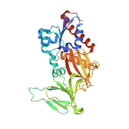
6N5C

Crystal structure of the catalytic domain of PPIP5K2 in complex with AMPPNP and 5-PCF2Am-InsP5


Experimental Data Snapshot

  • Method: X-RAY DIFFRACTION
  • Resolution: 1.95 Å
  • R-Value Free: 0.214 
  • R-Value Work: 0.175 
  • R-Value Observed: 0.178 

Starting Model: experimental
View more details

wwPDB Validation   3D Report Full Report


Ligand Structure Quality Assessment 


This is version 1.2 of the entry. See complete history


Literature

Synthesis of an alpha-phosphono-alpha , alpha-difluoroacetamide analogue of the diphosphoinositol pentakisphosphate 5-InsP7.

Riley, A.M.Wang, H.Shears, S.B.Potter, B.V.L.

(2019) Medchemcomm 10: 1165-1172

  • DOI: https://doi.org/10.1039/c9md00163h
  • Primary Citation of Related Structures:  
    6N5C

  • PubMed Abstract: 

    Diphosphoinositol phosphates (PP-InsPs) are an evolutionarily ancient group of signalling molecules that are essential to cellular and organismal homeostasis. As the detailed mechanisms of PP-InsP signalling begin to emerge, synthetic analogues of PP-InsPs containing stabilised mimics of the labile diphosphate group can provide valuable investigational tools. We synthesised 5-PCF 2 Am-InsP 5 ( 1 ), a novel fluorinated phosphonate analogue of 5-PP-InsP 5 , and obtained an X-ray crystal structure of 1 in complex with diphosphoinositol pentakisphosphate kinase 2 (PPIP5K2). 5-PCF 2 Am-InsP 5 binds to the kinase domain of PPIP5K2 in a similar orientation to that of the natural substrate 5-PP-InsP 5 and the PCF 2 Am structure can mimic many aspects of the diphosphate group in 5-PP-InsP 5 . We propose that 1 , the structural and electronic properties of which are in some ways complementary to those of existing phosphonoacetate and methylenebisphosphonate analogues of 5-PP-InsP 5 , may be a useful addition to the expanding array of chemical tools for the investigation of signalling by PP-InsPs. The PCF 2 Am group may also deserve attention for wider application as a diphosphate mimic.


  • Organizational Affiliation

    Medicinal Chemistry and Drug Discovery , Department of Pharmacology , University of Oxford , Mansfield Road , Oxford OX1 3QT , UK . Email: [email protected] ; ; Tel: +44 (0)1865 271945.


Macromolecules
Find similar proteins by:  (by identity cutoff)  |  3D Structure
Entity ID: 1
MoleculeChains Sequence LengthOrganismDetailsImage
Inositol hexakisphosphate and diphosphoinositol-pentakisphosphate kinase 2330Homo sapiensMutation(s): 0 
Gene Names: PPIP5K2HISPPD1KIAA0433VIP2
EC: 2.7.4.21 (PDB Primary Data), 2.7.4.24 (PDB Primary Data)
UniProt & NIH Common Fund Data Resources
Find proteins for O43314 (Homo sapiens)
Explore O43314 
Go to UniProtKB:  O43314
PHAROS:  O43314
GTEx:  ENSG00000145725 
Entity Groups  
Sequence Clusters30% Identity50% Identity70% Identity90% Identity95% Identity100% Identity
UniProt GroupO43314
Sequence Annotations
Expand
  • Reference Sequence
Small Molecules
Ligands 5 Unique
IDChains Name / Formula / InChI Key2D Diagram3D Interactions
KDJ
Query on KDJ

Download Ideal Coordinates CCD File 
G [auth A](1,1-difluoro-2-oxo-2-{[(1s,2R,3S,4s,5R,6S)-2,3,4,5,6-pentakis(phosphonooxy)cyclohexyl]amino}ethyl)phosphonic acid
C8 H19 F2 N O24 P6
XBHZOGSBYRIXJA-QWBQGLJISA-N
ANP
Query on ANP

Download Ideal Coordinates CCD File 
B [auth A]PHOSPHOAMINOPHOSPHONIC ACID-ADENYLATE ESTER
C10 H17 N6 O12 P3
PVKSNHVPLWYQGJ-KQYNXXCUSA-N
EDO
Query on EDO

Download Ideal Coordinates CCD File 
I [auth A],
K [auth A],
L [auth A],
M [auth A],
O [auth A]
1,2-ETHANEDIOL
C2 H6 O2
LYCAIKOWRPUZTN-UHFFFAOYSA-N
ACT
Query on ACT

Download Ideal Coordinates CCD File 
H [auth A],
J [auth A],
N [auth A]
ACETATE ION
C2 H3 O2
QTBSBXVTEAMEQO-UHFFFAOYSA-M
MG
Query on MG

Download Ideal Coordinates CCD File 
C [auth A],
D [auth A],
E [auth A],
F [auth A]
MAGNESIUM ION
Mg
JLVVSXFLKOJNIY-UHFFFAOYSA-N
Experimental Data & Validation

Experimental Data

  • Method: X-RAY DIFFRACTION
  • Resolution: 1.95 Å
  • R-Value Free: 0.214 
  • R-Value Work: 0.175 
  • R-Value Observed: 0.178 
  • Space Group: P 21 21 21
Unit Cell:
Length ( Å )Angle ( ˚ )
a = 88.604α = 90
b = 111.032β = 90
c = 41.429γ = 90
Software Package:
Software NamePurpose
REFMACrefinement
HKL-2000data reduction
HKL-2000data scaling
REFMACphasing

Structure Validation

View Full Validation Report


Ligand Structure Quality Assessment 


Entry History & Funding Information

Deposition Data


Funding OrganizationLocationGrant Number
National Institutes of Health/National Institute of Environmental Health Sciences (NIH/NIEHS)United States1ZIAES080046-29

Revision History  (Full details and data files)

  • Version 1.0: 2019-08-21
    Type: Initial release
  • Version 1.1: 2019-12-18
    Changes: Author supporting evidence
  • Version 1.2: 2023-10-11
    Changes: Data collection, Database references, Derived calculations, Refinement description