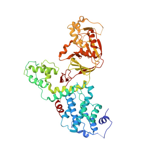
6NTW

Crystal structure of E. coli YcbB


Experimental Data Snapshot

  • Method: X-RAY DIFFRACTION
  • Resolution: 2.76 Å
  • R-Value Free: 0.298 
  • R-Value Work: 0.256 
  • R-Value Observed: 0.258 

wwPDB Validation   3D Report Full Report


Ligand Structure Quality Assessment 


This is version 1.3 of the entry. See complete history


Literature

Structural insight into YcbB-mediated beta-lactam resistance in Escherichia coli.

Caveney, N.A.Caballero, G.Voedts, H.Niciforovic, A.Worrall, L.J.Vuckovic, M.Fonvielle, M.Hugonnet, J.E.Arthur, M.Strynadka, N.C.J.

(2019) Nat Commun 10: 1849-1849

  • DOI: https://doi.org/10.1038/s41467-019-09507-0
  • Primary Citation of Related Structures:  
    6NTW, 6NTZ

  • PubMed Abstract: 

    The bacterial cell wall plays a crucial role in viability and is an important drug target. In Escherichia coli, the peptidoglycan crosslinking reaction to form the cell wall is primarily carried out by penicillin-binding proteins that catalyse D,D-transpeptidase activity. However, an alternate crosslinking mechanism involving the L,D-transpeptidase YcbB can lead to bypass of D,D-transpeptidation and beta-lactam resistance. Here, we show that the crystallographic structure of YcbB consists of a conserved L,D-transpeptidase catalytic domain decorated with a subdomain on the dynamic substrate capping loop, peptidoglycan-binding and large scaffolding domains. Meropenem acylation of YcbB gives insight into the mode of inhibition by carbapenems, the singular antibiotic class with significant activity against L,D-transpeptidases. We also report the structure of PBP5-meropenem to compare interactions mediating inhibition. Additionally, we probe the interaction network of this pathway and assay beta-lactam resistance in vivo. Our results provide structural insights into the mechanism of action and the inhibition of L,D-transpeptidation, and into YcbB-mediated antibiotic resistance.


  • Organizational Affiliation

    Department of Biochemistry and Molecular Biology and the Centre for Blood Research, University of British Columbia, Vancouver, V6T 1Z3, BC, Canada.


Macromolecules
Find similar proteins by:  (by identity cutoff)  |  3D Structure
Entity ID: 1
MoleculeChains Sequence LengthOrganismDetailsImage
Probable L,D-transpeptidase YcbB585Escherichia coli K-12Mutation(s): 0 
Gene Names: ycbBb0925JW0908
EC: 2
UniProt
Find proteins for P22525 (Escherichia coli (strain K12))
Explore P22525 
Go to UniProtKB:  P22525
Entity Groups  
Sequence Clusters30% Identity50% Identity70% Identity90% Identity95% Identity100% Identity
UniProt GroupP22525
Sequence Annotations
Expand
  • Reference Sequence
Experimental Data & Validation

Experimental Data

  • Method: X-RAY DIFFRACTION
  • Resolution: 2.76 Å
  • R-Value Free: 0.298 
  • R-Value Work: 0.256 
  • R-Value Observed: 0.258 
  • Space Group: P 43 21 2
Unit Cell:
Length ( Å )Angle ( ˚ )
a = 126.499α = 90
b = 126.499β = 90
c = 88.801γ = 90
Software Package:
Software NamePurpose
PHENIXrefinement
Aimlessdata scaling
PDB_EXTRACTdata extraction
XDSdata reduction
PHASERphasing

Structure Validation

View Full Validation Report


Ligand Structure Quality Assessment 


Entry History & Funding Information

Deposition Data


Funding OrganizationLocationGrant Number
Canadian Institutes of Health Research (CIHR)Canada--

Revision History  (Full details and data files)

  • Version 1.0: 2019-03-20
    Type: Initial release
  • Version 1.1: 2019-05-08
    Changes: Data collection, Database references
  • Version 1.2: 2020-01-08
    Changes: Author supporting evidence
  • Version 1.3: 2024-11-20
    Changes: Data collection, Database references, Derived calculations, Structure summary