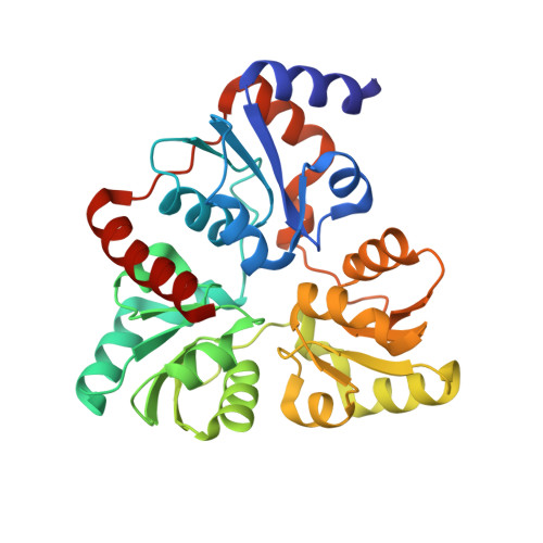
6ORA

An Unexpected Intermediate in the Reaction Catalyzed by Quinolinate Synthase


Experimental Data Snapshot

  • Method: X-RAY DIFFRACTION
  • Resolution: 2.20 Å
  • R-Value Free: 0.243 
  • R-Value Work: 0.196 
  • R-Value Observed: 0.200 

Starting Model: experimental
View more details

wwPDB Validation   3D Report Full Report


Ligand Structure Quality Assessment 


This is version 1.3 of the entry. See complete history


Literature

An Unexpected Species Determined by X-ray Crystallography that May Represent an Intermediate in the Reaction Catalyzed by Quinolinate Synthase.

Esakova, O.A.Silakov, A.Grove, T.L.Warui, D.M.Yennawar, N.H.Booker, S.J.

(2019) J Am Chem Soc 141: 14142-14151

  • DOI: https://doi.org/10.1021/jacs.9b02513
  • Primary Citation of Related Structures:  
    6NSO, 6NSU, 6OR8, 6ORA

  • PubMed Abstract: 

    Quinolinic acid is a common intermediate in the biosynthesis of nicotinamide adenine dinucleotide and its derivatives in all organisms that synthesize the molecule de novo. In most prokaryotes, it is formed from the condensation of dihydroxyacetone phosphate (DHAP) and iminoaspartate (IA) by the action of quinolinate synthase (NadA). NadA contains a [4Fe-4S] cluster cofactor with a unique noncysteinyl-ligated iron ion (Fe a ), which is proposed to bind the hydroxyl group of an intermediate in its reaction to facilitate a dehydration step. However, direct evidence for this role in catalysis has yet to be provided, and the exact chemical mechanism that underlies this transformation remains elusive. Herein, we present a structure of NadA from Pyrococcus horikoshii (PhNadA) in complex with IA and show that a carboxylate group of the molecule is ligated to Fe a of the iron-sulfur cluster, occupying the site to which DHAP has been proposed to bind during catalysis. When crystals of PhNadA in complex with IA are soaked briefly in DHAP before freezing, electron density for a new molecule is observed, which we suggest is related to an intermediate in the reaction. Similar, but slightly different, "intermediates" are observed when crystals of a PhNadA Glu198Gln variant are incubated with DHAP, oxaloacetate, and ammonium chloride, conditions under which IA is formed chemically. Continuous-wave and pulse electron paramagnetic resonance techniques are used to verify the binding mode of substrates and proposed intermediates in frozen solution.


Macromolecules
Find similar proteins by:  (by identity cutoff)  |  3D Structure
Entity ID: 1
MoleculeChains Sequence LengthOrganismDetailsImage
Quinolinate synthase A
A, B
300Pyrococcus horikoshii OT3Mutation(s): 1 
Gene Names: nadAPH0013
EC: 2.5.1.72
UniProt
Find proteins for O57767 (Pyrococcus horikoshii (strain ATCC 700860 / DSM 12428 / JCM 9974 / NBRC 100139 / OT-3))
Explore O57767 
Go to UniProtKB:  O57767
Entity Groups  
Sequence Clusters30% Identity50% Identity70% Identity90% Identity95% Identity100% Identity
UniProt GroupO57767
Sequence Annotations
Expand
  • Reference Sequence
Small Molecules
Ligands 5 Unique
IDChains Name / Formula / InChI Key2D Diagram3D Interactions
SF4
Query on SF4

Download Ideal Coordinates CCD File 
C [auth A],
G [auth B]
IRON/SULFUR CLUSTER
Fe4 S4
LJBDFODJNLIPKO-UHFFFAOYSA-N
5XR (Subject of Investigation/LOI)
Query on 5XR

Download Ideal Coordinates CCD File 
I [auth B]2-hydroxy-N-[(1S)-1-hydroxy-3-oxopropyl]-L-aspartic acid
C7 H11 N O7
CVFQCYNIQPQUNC-MHTLYPKNSA-N
5XW (Subject of Investigation/LOI)
Query on 5XW

Download Ideal Coordinates CCD File 
E [auth A](2~{Z})-2-(1-oxidanyl-3-oxidanylidene-propyl)iminobutanedioic acid
C7 H9 N O6
BKMLHLNYNPUNII-RZTYMHDWSA-N
ACT
Query on ACT

Download Ideal Coordinates CCD File 
D [auth A],
H [auth B]
ACETATE ION
C2 H3 O2
QTBSBXVTEAMEQO-UHFFFAOYSA-M
CL
Query on CL

Download Ideal Coordinates CCD File 
F [auth A],
J [auth B]
CHLORIDE ION
Cl
VEXZGXHMUGYJMC-UHFFFAOYSA-M
Experimental Data & Validation

Experimental Data

  • Method: X-RAY DIFFRACTION
  • Resolution: 2.20 Å
  • R-Value Free: 0.243 
  • R-Value Work: 0.196 
  • R-Value Observed: 0.200 
  • Space Group: P 1 21 1
Unit Cell:
Length ( Å )Angle ( ˚ )
a = 53.052α = 90
b = 52.134β = 96.27
c = 97.803γ = 90
Software Package:
Software NamePurpose
PHENIXrefinement
HKL-2000data reduction
HKL-2000data scaling
PHASERphasing

Structure Validation

View Full Validation Report


Ligand Structure Quality Assessment 


Entry History & Funding Information

Deposition Data


Funding OrganizationLocationGrant Number
National Institutes of Health/National Human Genome Research Institute (NIH/NHGRI)United StatesGM - 122595
National Science Foundation (NSF, United States)United StatesMCB - 1716686
National Science Foundation (NSF, United States)United StatesCHE - 1659679
Howard Hughes Medical Institute (HHMI)United States--

Revision History  (Full details and data files)

  • Version 1.0: 2019-08-21
    Type: Initial release
  • Version 1.1: 2019-09-25
    Changes: Data collection, Database references
  • Version 1.2: 2019-11-20
    Changes: Author supporting evidence
  • Version 1.3: 2023-10-11
    Changes: Data collection, Database references, Derived calculations, Refinement description