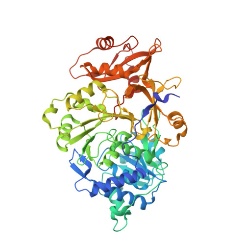
6Q2M

Crystal structure of Photinus pyralis Luciferase Pps6 mutant in complex with DLSA


Experimental Data Snapshot

  • Method: X-RAY DIFFRACTION
  • Resolution: 2.75 Å
  • R-Value Free: 0.218 
  • R-Value Work: 0.194 
  • R-Value Observed: 0.195 

Starting Model: experimental
View more details

wwPDB Validation   3D Report Full Report


Ligand Structure Quality Assessment 


This is version 1.4 of the entry. See complete history


Literature

Mutagenesis and Structural Studies Reveal the Basis for the Activity and Stability Properties That Distinguish thePhotinusLuciferasesscintillansandpyralis.

Branchini, B.R.Fontaine, D.M.Southworth, T.L.Huta, B.P.Racela, A.Patel, K.D.Gulick, A.M.

(2019) Biochemistry 58: 4293-4303

  • DOI: https://doi.org/10.1021/acs.biochem.9b00719
  • Primary Citation of Related Structures:  
    6Q2M

  • PubMed Abstract: 

    The dazzling yellow-green light emission of the common North American firefly Photinus pyralis and other bioluminescent organisms has provided a wide variety of prominent research applications like reporter gene assays and in vivo imaging methods. While the P. pyralis enzyme has been extensively studied, only recently has a second Photinus luciferase been cloned from the species scintillans . Even though the enzymes share very high sequence identity (89.8%), the color of the light they emit, their specific activity and their stability to heat, pH, and chemical denaturation are quite different with the scintillans luciferase being generally more resistant. Through the construction and evaluation of the properties of chimeric domain swapped, single point, and various combined variants, we have determined that only six amino acid changes are necessary to confer all of the properties of the scintillans enzyme to wild-type P. pyralis luciferase. Altered stability properties were attributed to four of the amino acid changes (T214N/S276T/H332N/E354N), and single mutations each predominantly changed emission color (Y255F) and specific activity (A222C). Results of a crystallographic study of the P. pyralis enzyme containing the six changes (Pps6) provide some insight into the structural basis for some of the documented property differences.


  • Organizational Affiliation

    Department of Chemistry , Connecticut College , New London , Connecticut 06320 , United States.


Macromolecules
Find similar proteins by:  (by identity cutoff)  |  3D Structure
Entity ID: 1
MoleculeChains Sequence LengthOrganismDetailsImage
Luciferin 4-monooxygenase
A, B, C
555Photinus pyralisMutation(s): 6 
EC: 1.13.12.7
UniProt
Find proteins for P08659 (Photinus pyralis)
Explore P08659 
Go to UniProtKB:  P08659
Entity Groups  
Sequence Clusters30% Identity50% Identity70% Identity90% Identity95% Identity100% Identity
UniProt GroupP08659
Sequence Annotations
Expand
  • Reference Sequence
Small Molecules
Ligands 4 Unique
IDChains Name / Formula / InChI Key2D Diagram3D Interactions
SLU (Subject of Investigation/LOI)
Query on SLU

Download Ideal Coordinates CCD File 
CB [auth C],
HA [auth B],
W [auth A]
5'-O-[N-(DEHYDROLUCIFERYL)-SULFAMOYL] ADENOSINE
C21 H18 N8 O8 S3
LJLYTUYNVSHXQB-SOONXTGKSA-N
DYD
Query on DYD

Download Ideal Coordinates CCD File 
E [auth A],
F [auth A],
KA [auth C],
LA [auth C]
(2S,5S)-hexane-2,5-diol
C6 H14 O2
OHMBHFSEKCCCBW-WDSKDSINSA-N
SO4
Query on SO4

Download Ideal Coordinates CCD File 
D [auth A]
IA [auth C]
JA [auth C]
X [auth B]
Y [auth B]
D [auth A],
IA [auth C],
JA [auth C],
X [auth B],
Y [auth B],
Z [auth B]
SULFATE ION
O4 S
QAOWNCQODCNURD-UHFFFAOYSA-L
EDO
Query on EDO

Download Ideal Coordinates CCD File 
AA [auth B]
AB [auth C]
BA [auth B]
BB [auth C]
CA [auth B]
AA [auth B],
AB [auth C],
BA [auth B],
BB [auth C],
CA [auth B],
DA [auth B],
EA [auth B],
FA [auth B],
G [auth A],
GA [auth B],
H [auth A],
I [auth A],
J [auth A],
K [auth A],
L [auth A],
M [auth A],
MA [auth C],
N [auth A],
NA [auth C],
O [auth A],
OA [auth C],
P [auth A],
PA [auth C],
Q [auth A],
QA [auth C],
R [auth A],
RA [auth C],
S [auth A],
SA [auth C],
T [auth A],
TA [auth C],
U [auth A],
UA [auth C],
V [auth A],
VA [auth C],
WA [auth C],
XA [auth C],
YA [auth C],
ZA [auth C]
1,2-ETHANEDIOL
C2 H6 O2
LYCAIKOWRPUZTN-UHFFFAOYSA-N
Experimental Data & Validation

Experimental Data

  • Method: X-RAY DIFFRACTION
  • Resolution: 2.75 Å
  • R-Value Free: 0.218 
  • R-Value Work: 0.194 
  • R-Value Observed: 0.195 
  • Space Group: P 1 21 1
Unit Cell:
Length ( Å )Angle ( ˚ )
a = 66.268α = 90
b = 95.455β = 101.556
c = 152.02γ = 90
Software Package:
Software NamePurpose
PHENIXrefinement
XDSdata reduction
autoPROCdata scaling
PHASERphasing

Structure Validation

View Full Validation Report


Ligand Structure Quality Assessment 


Entry History & Funding Information

Deposition Data


Funding OrganizationLocationGrant Number
National Institutes of Health/National Human Genome Research Institute (NIH/NHGRI)United StatesR01GM116957

Revision History  (Full details and data files)

  • Version 1.0: 2019-10-16
    Type: Initial release
  • Version 1.1: 2019-10-23
    Changes: Data collection, Database references
  • Version 1.2: 2019-11-06
    Changes: Data collection, Database references
  • Version 1.3: 2019-12-18
    Changes: Author supporting evidence
  • Version 1.4: 2023-10-11
    Changes: Data collection, Database references, Refinement description