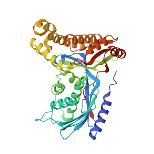
6QJE

Structure of human galactokinase 1 bound with 4-{[2-(Methylsulfonyl)-1H-imidazol-1-yl]methyl}-1,3-thiazole


Experimental Data Snapshot

  • Method: X-RAY DIFFRACTION
  • Resolution: 2.40 Å
  • R-Value Free: 0.269 
  • R-Value Work: 0.217 
  • R-Value Observed: 0.220 

Starting Model: experimental
View more details

wwPDB Validation   3D Report Full Report


Ligand Structure Quality Assessment 


This is version 1.3 of the entry. See complete history


Literature

Structure of human galactokinase 1 bound with 4-{[2-(Methylsulfonyl)-1H-imidazol-1-yl]methyl}-1,3-thiazole

Mackinnon, S.R.Bezerra, G.A.Zhang, M.Foster, W.Krojer, T.Brandao-Neto, J.Douangamath, A.Arrowsmith, C.Edwards, A.Bountra, C.Brennan, P.Lai, K.Yue, W.W.

To be published.

Macromolecules
Find similar proteins by:  (by identity cutoff)  |  3D Structure
Entity ID: 1
MoleculeChains Sequence LengthOrganismDetailsImage
Galactokinase
A, B, C, D
392Homo sapiensMutation(s): 2 
Gene Names: GALK1GALK
EC: 2.7.1.6
UniProt & NIH Common Fund Data Resources
Find proteins for P51570 (Homo sapiens)
Explore P51570 
Go to UniProtKB:  P51570
PHAROS:  P51570
GTEx:  ENSG00000108479 
Entity Groups  
Sequence Clusters30% Identity50% Identity70% Identity90% Identity95% Identity100% Identity
UniProt GroupP51570
Sequence Annotations
Expand
  • Reference Sequence
Small Molecules
Ligands 4 Unique
IDChains Name / Formula / InChI Key2D Diagram3D Interactions
HFK
Query on HFK

Download Ideal Coordinates CCD File 
F [auth A],
I [auth B],
L [auth C]
2-(1,3-benzoxazol-2-ylamino)spiro[1,6,7,8-tetrahydroquinazoline-4,1'-cyclohexane]-5-one
C20 H22 N4 O2
AZBGHUMVHJKFOO-UHFFFAOYSA-N
J4Q
Query on J4Q

Download Ideal Coordinates CCD File 
G [auth A],
J [auth B],
N [auth D]
4-[(2-methylsulfonylimidazol-1-yl)methyl]-1,3-thiazole
C8 H9 N3 O2 S2
DWPGGNJRLYKHCD-UHFFFAOYSA-N
GAL
Query on GAL

Download Ideal Coordinates CCD File 
E [auth A],
H [auth B],
K [auth C]
beta-D-galactopyranose
C6 H12 O6
WQZGKKKJIJFFOK-FPRJBGLDSA-N
SO4
Query on SO4

Download Ideal Coordinates CCD File 
M [auth C]SULFATE ION
O4 S
QAOWNCQODCNURD-UHFFFAOYSA-L
Experimental Data & Validation

Experimental Data

  • Method: X-RAY DIFFRACTION
  • Resolution: 2.40 Å
  • R-Value Free: 0.269 
  • R-Value Work: 0.217 
  • R-Value Observed: 0.220 
  • Space Group: P 1 21 1
Unit Cell:
Length ( Å )Angle ( ˚ )
a = 73.703α = 90
b = 114.873β = 100.56
c = 121.044γ = 90
Software Package:
Software NamePurpose
Aimlessdata scaling
PHASERphasing
PHENIXrefinement
PDB_EXTRACTdata extraction
XDSdata reduction

Structure Validation

View Full Validation Report


Ligand Structure Quality Assessment 


Entry History & Funding Information

Deposition Data


Funding OrganizationLocationGrant Number
Wellcome TrustUnited Kingdom092809/Z/10/Z

Revision History  (Full details and data files)

  • Version 1.0: 2019-02-27
    Type: Initial release
  • Version 1.1: 2020-07-29
    Type: Remediation
    Reason: Carbohydrate remediation
    Changes: Data collection, Derived calculations, Structure summary
  • Version 1.2: 2024-01-24
    Changes: Data collection, Database references, Refinement description, Structure summary
  • Version 1.3: 2024-11-06
    Changes: Structure summary